Explodable 3D Dog Skull for Veterinary Education
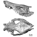
3D models of Ocnotherium skull
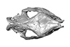
3D models of Kalakocetus, the earliest Cetacea
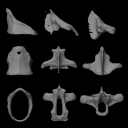
3D GM dataset of bird skeletal variation
Skeletal embryonic development in the catshark
Bony connexions of the petrosal bone of extant hippos
bony labyrinth (14) , inner ear (11) , geometric morphometrics (10) , CT-scan (10) , Eocene (10) , Micro-CT (9) , Miocene (8)
Lionel Hautier (24) , Maëva Judith Orliac (23) , Laurent Marivaux (18) , Renaud Lebrun (14) , Rodolphe Tabuce (14) , Pierre-Olivier Antoine (13) , Bastien Mennecart (13)
|
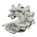
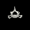
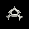
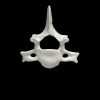
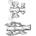
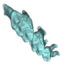
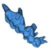
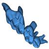
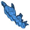
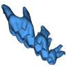
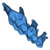
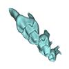
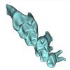
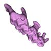
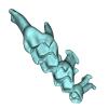
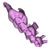
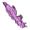
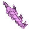
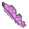
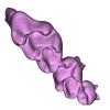
3D models related to the publication: “The functional significance of aberrant cervical counts in sloths: insights from automated exhaustive analysis of cervical range of motion”Luisa J. Merten
Published online: 04/11/2023 |
|
M3#1260cervical vertebral series (7 vertebrae) Type: "3D_surfaces"doi: 10.18563/m3.sf.1260 state:published |
Download 3D surface file |
Bradypus variegatus ZMB_Mam_91345 View specimen
|
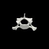
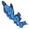
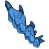
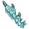
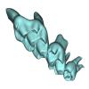
M3#1261cervical vertebral series (8 vertebrae) + first thoracic vertebra Type: "3D_surfaces"doi: 10.18563/m3.sf.1261 state:published |
Download 3D surface file |
Bradypus variegatus ZMB_Mam_35824 View specimen

|
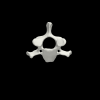
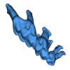
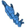
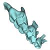
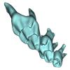
M3#1262cervical vertebral series (8 vertebrae) + first & second thoracic vertebra Type: "3D_surfaces"doi: 10.18563/m3.sf.1262 state:published |
Download 3D surface file |
Choloepus didactylus ZMB_Mam_38388 View specimen
|
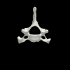
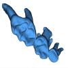
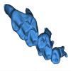
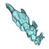
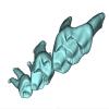
M3#1263cervical vertebral series (7 vertebrae) Type: "3D_surfaces"doi: 10.18563/m3.sf.1263 state:published |
Download 3D surface file |
Choloepus didactylus ZMB_Mam_102634 View specimen
|
M3#1264cervical vertebral series (6 vertebrae) + first thoracic vertebra Type: "3D_surfaces"doi: 10.18563/m3.sf.1264 state:published |
Download 3D surface file |
Tamandua tetradactyla ZMB_Mam_91288 View specimen
|
M3#1266cervical vertebral series (7 vertebrae) + first thoracic vertebra Type: "3D_surfaces"doi: 10.18563/m3.sf.1266 state:published |
Download 3D surface file |
Glossotherium robustum MNHN_n/n View specimen
|
M3#1267cervical vertebral series (7 vertebrae) + first thoracic vertebra Type: "3D_surfaces"doi: 10.18563/m3.sf.1267 state:published |
Download 3D surface file |
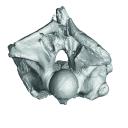
The present 3D Dataset contains the 3D models of the sacral vertebrae analyzed in “Sacral co-ossification in dinosaurs: The oldest record of fused sacral vertebrae in Dinosauria and the diversity of sacral co-ossification patterns in the group”.
Buriolestes schultzi CAPPA/UFSM 0035 View specimen

|
M3#705Sacral vertebrae of Buriolestes schultzi Type: "3D_surfaces"doi: 10.18563/m3.sf.705 state:published |
Download 3D surface file |
indet indet CAPPA/UFSM 0228 View specimen
|
M3#706Sacral vertebrae of a saurischian dinosaur indet. Type: "3D_surfaces"doi: 10.18563/m3.sf.706 state:published |
Download 3D surface file |
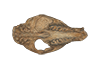
The present 3D Dataset contains the 3D model analyzed in the article : Dubied et al. (2021), Endocranium and ecology of Eurotherium theriodis, a European hyaenodont mammal from the Lutetian. Acta Palaeontologica Polonica 2021, https://doi.org/10.4202/app.00771.2020
Eurotherium theriodis NMB.Em12 View specimen
|
M3#381NMB.Em12 unprepared specimen Type: "3D_surfaces"doi: 10.18563/m3.sf.381 state:published |
Download 3D surface file |
|
M3#382NMB.Em12 cranium Type: "3D_surfaces"doi: 10.18563/m3.sf.382 state:published |
Download 3D surface file |
|
M3#383NMB.Em12 endocast Type: "3D_surfaces"doi: 10.18563/m3.sf.383 state:published |
Download 3D surface file |

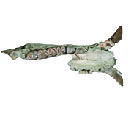
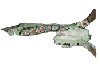
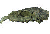
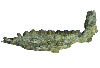
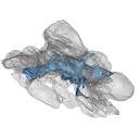
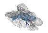
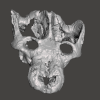
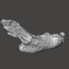
The present 3D Dataset contains the 3D models analyzed in: Perrichon et al., 2023. Neuroanatomy and pneumaticity of Voay robustus and its implications for crocodylid phylogeny and palaeoecology.
Crocodylus niloticus MHNL 50001387 View specimen
|
M3#1202Skull, inner ear, pharyngotympanic sinus and neurovascular system Type: "3D_surfaces"doi: 10.18563/m3.sf.1202 state:published |
Download 3D surface file |
Voay robustus MNHN F.1908-5 View specimen
|
M3#1203Skull, inner ear, pharyngotympanic sinus and neurovascular system Type: "3D_surfaces"doi: 10.18563/m3.sf.1203 state:published |
Download 3D surface file |
Voay robustus NHMUK PV R 36684 View specimen
|
M3#1204Skull, inner ear, pharyngotympanic sinus and neurovascular system Type: "3D_surfaces"doi: 10.18563/m3.sf.1204 state:published |
Download 3D surface file |
Voay robustus NHMUK PV R 36685 View specimen
|
M3#1205Skull, inner ear, pharyngotympanic sinus and neurovascular system Type: "3D_surfaces"doi: 10.18563/m3.sf.1205 state:published |
Download 3D surface file |
Osteolaemus tetraspis UCBLZ 2019-1-236 View specimen
|
M3#1208Skull, inner ear, pharyngotympanic sinus and neurovascular system Type: "3D_surfaces"doi: 10.18563/m3.sf.1208 state:published |
Download 3D surface file |
Mecistops sp. UM N89 View specimen
|
M3#1207Skull, inner ear, pharyngotympanic sinus and neurovascular system Type: "3D_surfaces"doi: 10.18563/m3.sf.1207 state:published |
Download 3D surface file |
Voay robustus NHMUK PV R 2204 View specimen
|
M3#1206Skull, inner ear, pharyngotympanic sinus, intertympanic sinus and neurovascular system Type: "3D_surfaces"doi: 10.18563/m3.sf.1206 state:published |
Download 3D surface file |
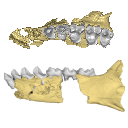
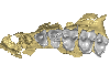
This contribution contains the 3D models described and figured in: New remains of Neotropical bunodont litopterns and the systematics of Megadolodinae (Mammalia: Litopterna). Geodiversitas.
Megadolodus molariformes VPPLT 974 View specimen
|
M3#1020Partial mandible with the symphysis and left body, bearing the alveoli of ?i2, right and left ?i3, alveolus of right c and p1, roots of left p1, and the left p2–m3 of Megadolodus molariformes (Litopterna, Mammalia) Type: "3D_surfaces"doi: 10.18563/m3.sf.1020 state:published |
Download 3D surface file |
Neodolodus colombianus VPPLT 1696 View specimen
|
M3#1021Almost complete skull with left and right ?I2 and P1–M3 Type: "3D_surfaces"doi: 10.18563/m3.sf.1021 state:published |
Download 3D surface file |
|
M3#1022Partial mandible with complete right and left dentition except for left ?i2 Type: "3D_surfaces"doi: 10.18563/m3.sf.1022 state:published |
Download 3D surface file |
This contribution contains the 3D models described and figured in the following publication: Paulina-Carabajal, A. and Nieto, M. N. In press. Brief comment on the brain and inner ear of Giganotosaurus carolinii (Dinosauria: Theropoda) based on CT scans. Ameghiniana. https://doi.org/10.5710/AMGH.25.10.2019.3237
Giganotosaurus carolinii MUCPv-CH-1 View specimen
|
M3#504The current file contents 3D models of the braincase, brain, left and right inner ears Type: "3D_surfaces"doi: 10.18563/m3.sf.504 state:published |
Download 3D surface file |
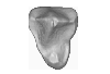
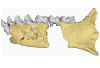
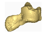
This contribution contains the 3D models of the fossil remains (maxilla, dentary, and talus) attributed to Djebelemur martinezi, a ca. 50 Ma primate from Tunisia (Djebel Chambi), described and figured in the following publication: Marivaux et al. (2013), Djebelemur, a tiny pre-tooth-combed primate from the Eocene of Tunisia: a glimpse into the origin of crown strepsirhines. PLoS ONE. https://doi.org/10.1371/journal.pone.0080778
Djebelemur martinezi CBI-1-544 View specimen
|
M3#365CBI-1-544, left maxilla preserving P3-M3 and alveoli for P2 and C1 Type: "3D_surfaces"doi: 10.18563/m3.sf.365 state:published |
Download 3D surface file |
Djebelemur martinezi CBI-1-567 View specimen
|
M3#363Isolated left upper P4 Type: "3D_surfaces"doi: 10.18563/m3.sf.363 state:published |
Download 3D surface file |
Djebelemur martinezi CBI-1-565-577-587-580 View specimen
|
M3#366- CBI-1-565, a damaged right mandible, which consists of three isolated pieces found together and reassembled here: the anterior part of the dentary bears the p3 and m1, and alveoli for p4, p2 and c, while the posterior part preserves m3 and a portion of the ascending ramus; the m2 was found isolated but in the same small calcareous block treated by acid processing. - CBI-1-577, isolated right lower p4. - CBI-1-587, isolated left lower p2 (reversed). - CBI-1-580, isolated left lower canine (reversed). Type: "3D_surfaces"doi: 10.18563/m3.sf.366 state:published |
Download 3D surface file |
Djebelemur martinezi CBI-1-545 View specimen
|
M3#364Right Talus Type: "3D_surfaces"doi: 10.18563/m3.sf.364 state:published |
Download 3D surface file |
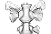
The present 3D Dataset contains the 3D models of a skull and lower jaw of the holotype of Santagnathus mariensis, described in “Old fossil findings in the Upper Triassic rocks of southern Brazil improve diversity of traversodontid cynodonts (Therapsida, Cynodontia)”
Santagnathus mariensis UFRGS-PV-1419-T View specimen
|
M3#1157Skull Type: "3D_surfaces"doi: 10.18563/m3.sf.1157 state:published |
Download 3D surface file |
|
M3#1158Lower jaw Type: "3D_surfaces"doi: 10.18563/m3.sf.1158 state:published |
Download 3D surface file |
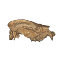
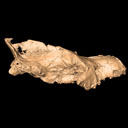
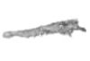
The present 3D Dataset contains the 3D model of a specimen of Metamynodon planifrons (UNISTRA.2015.0.1106) described and figured in: Veine-Tonizzo, L., Tissier, J., Bukhsianidze, M., Vasilyan, D., Becker, D., 2023, Cranial morphology and phylogenetic relationships of Amynodontidae Scott & Osborn, 1883 (Perissodactyla, Rhinocerotoidea).
Metamynodon planifrons UNISTRA.2015.0.1106 View specimen
|
M3#716Textured 3D surface model of the skull of the specimen UNISTRA.2015.0.1106 with right C1 and both rows of P2-M3. Type: "3D_surfaces"doi: 10.18563/m3.sf.716 state:published |
Download 3D surface file |
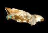
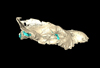
This contribution provides for the first time the 3D model of the type specimen of Molassitherium delemontense (Mammalia, Rhinocerotidae) described in the following publication: Becker et al. (2013), Journal of Systematic Palaeontology, Vol. 11, Issue 8, 947–972, https://doi.org/10.1080/14772019.2012.699007. Conservation issues of the specimen and solutions using 3D model and 3D prints are detailed.
Molassitherium delemontense MJSN POI007–245 View specimen
|
M3#384Skull of Molassitherium delemontense Becker and Antoine, 2013 (in Becker et al. 2013): holotype Type: "3D_surfaces"doi: 10.18563/m3.sf.384 state:published |
Download 3D surface file |
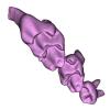
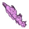
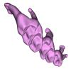
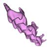
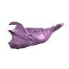
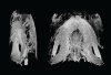
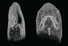
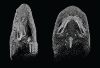
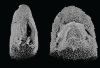
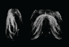
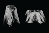
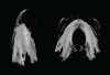
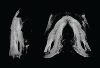
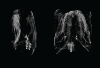
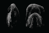
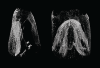
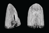
The present 3D Dataset contains the 3D models analyzed in Brualla et al., 2024: Comparative anatomy of the vocal apparatus in bats and implication for the diversity of laryngeal echolocation. Zoological Journal of the Linnean Society, vol. zlad180. (https://doi.org/10.1093/zoolinnean/zlad180). Bat larynges are understudied in the previous anatomical studies. The description and comparison of the different morphological traits might provide important proxies to investigate the evolutionary origin of laryngeal echolocation in bats.
Eonycteris spelaea VN18-026 View specimen

|
M3#1305Laryngeal cartilages and muscles of the cave nectar bat Type: "3D_surfaces"doi: 10.18563/m3.sf.1305 state:published |
Download 3D surface file |
Macroglossus sobrinus VN15-017 View specimen
|
M3#1306Laryngeal anatomy of Macroglossus sobrinus Type: "3D_surfaces"doi: 10.18563/m3.sf.1306 state:published |
Download 3D surface file |
Aselliscus dongbacana VTTu15-013 View specimen
|
M3#1307Laryngeal anatomy of Aselliscus dongbacana Type: "3D_surfaces"doi: 10.18563/m3.sf.1307 state:published |
Download 3D surface file |
Coelops frithii VN19-196 View specimen
|
M3#1308Laryngeal anatomy of Coelops frithii Type: "3D_surfaces"doi: 10.18563/m3.sf.1308 state:published |
Download 3D surface file |
Hipposideros larvatus VN18-209 View specimen
|
M3#1309Laryngeal anatomy of Hipposideros larvatus Type: "3D_surfaces"doi: 10.18563/m3.sf.1309 state:published |
Download 3D surface file |
Rhinolophus cornutus JP21-025 View specimen
|
M3#14763D surfaces of Rhinolophus cornutus Type: "3D_surfaces"doi: 10.18563/m3.sf.1476 state:published |
Download 3D surface file |
Rhinolophus macrotis VN11-089 View specimen
|
M3#1477Laryngeal cartilages and muscles of Rhinolophus macrotis Type: "3D_surfaces"doi: 10.18563/m3.sf.1477 state:published |
Download 3D surface file |
Lyroderma lyra VN17-535 View specimen
|
M3#1312Laryngeal anatomy of Lyroderma lyra Type: "3D_surfaces"doi: 10.18563/m3.sf.1312 state:published |
Download 3D surface file |
Saccolaimus mixtus A3257 View specimen
|
M3#1478Laryngeal components of Saccolaimus mixtus Type: "3D_surfaces"doi: 10.18563/m3.sf.1478 state:published |
Download 3D surface file |
Taphozous melanopogon VN17-0252 View specimen
|
M3#1479Laryngeal cartilages and muscles of Taphozous melanopogon Type: "3D_surfaces"doi: 10.18563/m3.sf.1479 state:published |
Download 3D surface file |
Artibeus jamaicensis AJ001 View specimen
|
M3#1316Laryngeal anatomy of Artibeus jamaicensis Type: "3D_surfaces"doi: 10.18563/m3.sf.1316 state:published |
Download 3D surface file |
Kerivoula hardwickii VN11-0043 View specimen
|
M3#1317Laryngeal anatomy of Kerivoula hardwickii Type: "3D_surfaces"doi: 10.18563/m3.sf.1317 state:published |
Download 3D surface file |
Myotis ater VN19-016 View specimen

|
M3#1318Laryngeal anatomy of Myotis ater Type: "3D_surfaces"doi: 10.18563/m3.sf.1318 state:published |
Download 3D surface file |
Myotis siligorensis VTTu14-018 View specimen
|
M3#1319Laryngeal anatomy of Myotis siligorensis Type: "3D_surfaces"doi: 10.18563/m3.sf.1319 state:published |
Download 3D surface file |
Suncus murinus KATS_835A View specimen
|
M3#1395Laryngeal anatomy of Suncus murinus Type: "3D_surfaces"doi: 10.18563/m3.sf.1395 state:published |
Download 3D surface file |
This contribution contains 3D models of upper molar rows of house mice (Mus musculus domesticus) belonging to Western European commensal and Sub-Antarctic feral populations. These two groups are characterized by different patterns of wear and alignment of the three molars along the row, related to contrasted masticatory demand in relation with their diet. These models are analyzed in the following publication: Renaud et al 2023, “Molar wear in house mice, insight into diet preferences at an ecological time scale?”, https://doi.org/10.1093/biolinnean/blad091
Mus musculus G09_06 View specimen
|
M3#1166right upper molar row Type: "3D_surfaces"doi: 10.18563/m3.sf.1166 state:published |
Download 3D surface file |
Mus musculus G09_10 View specimen
|
M3#1168right upper molar row Type: "3D_surfaces"doi: 10.18563/m3.sf.1168 state:published |
Download 3D surface file |
Mus musculus G09_15 View specimen
|
M3#1169right upper molar row Type: "3D_surfaces"doi: 10.18563/m3.sf.1169 state:published |
Download 3D surface file |
Mus musculus G09_16 View specimen
|
M3#1170right upper molar row Type: "3D_surfaces"doi: 10.18563/m3.sf.1170 state:published |
Download 3D surface file |
Mus musculus G09_17 View specimen
|
M3#1171right upper molar row Type: "3D_surfaces"doi: 10.18563/m3.sf.1171 state:published |
Download 3D surface file |
Mus musculus G09_21 View specimen
|
M3#1172right upper molar row Type: "3D_surfaces"doi: 10.18563/m3.sf.1172 state:published |
Download 3D surface file |
Mus musculus G09_26 View specimen
|
M3#1173right upper molar row Type: "3D_surfaces"doi: 10.18563/m3.sf.1173 state:published |
Download 3D surface file |
Mus musculus G09_27 View specimen
|
M3#1174right upper molar row Type: "3D_surfaces"doi: 10.18563/m3.sf.1174 state:published |
Download 3D surface file |
Mus musculus G09_29 View specimen
|
M3#1175right upper molar row Type: "3D_surfaces"doi: 10.18563/m3.sf.1175 state:published |
Download 3D surface file |
Mus musculus G09_65 View specimen
|
M3#1176right upper molar row Type: "3D_surfaces"doi: 10.18563/m3.sf.1176 state:published |
Download 3D surface file |
Mus musculus G09_66 View specimen
|
M3#1177right upper molar row Type: "3D_surfaces"doi: 10.18563/m3.sf.1177 state:published |
Download 3D surface file |
Mus musculus G93_03 View specimen
|
M3#1178right upper molar row Type: "3D_surfaces"doi: 10.18563/m3.sf.1178 state:published |
Download 3D surface file |
Mus musculus G93_04 View specimen
|
M3#1179right upper molar row Type: "3D_surfaces"doi: 10.18563/m3.sf.1179 state:published |
Download 3D surface file |
Mus musculus G93_10 View specimen
|
M3#1180right upper molar row Type: "3D_surfaces"doi: 10.18563/m3.sf.1180 state:published |
Download 3D surface file |
Mus musculus G93_11 View specimen
|
M3#1181right upper molar row Type: "3D_surfaces"doi: 10.18563/m3.sf.1181 state:published |
Download 3D surface file |
Mus musculus G93_13 View specimen
|
M3#1182right upper molar row Type: "3D_surfaces"doi: 10.18563/m3.sf.1182 state:published |
Download 3D surface file |
Mus musculus G93_14 View specimen
|
M3#1183right upper molar row Type: "3D_surfaces"doi: 10.18563/m3.sf.1183 state:published |
Download 3D surface file |
Mus musculus G93_15 View specimen
|
M3#1184right upper molar row Type: "3D_surfaces"doi: 10.18563/m3.sf.1184 state:published |
Download 3D surface file |
Mus musculus G93_24 View specimen
|
M3#1185left molar row Type: "3D_surfaces"doi: 10.18563/m3.sf.1185 state:published |
Download 3D surface file |
Mus musculus Tourch_7819 View specimen
|
M3#1186right upper molar row Type: "3D_surfaces"doi: 10.18563/m3.sf.1186 state:published |
Download 3D surface file |
Mus musculus G93_25 View specimen
|
M3#1187right upper molar row Type: "3D_surfaces"doi: 10.18563/m3.sf.1187 state:published |
Download 3D surface file |
Mus musculus Tourch_7821 View specimen
|
M3#1188right upper molar row Type: "3D_surfaces"doi: 10.18563/m3.sf.1188 state:published |
Download 3D surface file |
Mus musculus Tourch_7839 View specimen
|
M3#1189right upper molar row Type: "3D_surfaces"doi: 10.18563/m3.sf.1189 state:published |
Download 3D surface file |
Mus musculus Tourch_7873 View specimen
|
M3#1190right upper molar row Type: "3D_surfaces"doi: 10.18563/m3.sf.1190 state:published |
Download 3D surface file |
Mus musculus Tourch_7877 View specimen
|
M3#1196right upper molar row Type: "3D_surfaces"doi: 10.18563/m3.sf.1196 state:published |
Download 3D surface file |
Mus musculus Tourch_7922 View specimen
|
M3#1191right upper molar row Type: "3D_surfaces"doi: 10.18563/m3.sf.1191 state:published |
Download 3D surface file |
Mus musculus Tourch_7923 View specimen
|
M3#1192right upper molar row Type: "3D_surfaces"doi: 10.18563/m3.sf.1192 state:published |
Download 3D surface file |
Mus musculus Tourch_7925 View specimen
|
M3#1193right upper molar row Type: "3D_surfaces"doi: 10.18563/m3.sf.1193 state:published |
Download 3D surface file |
Mus musculus Tourch_7927 View specimen
|
M3#1194right upper molar row Type: "3D_surfaces"doi: 10.18563/m3.sf.1194 state:published |
Download 3D surface file |
Mus musculus Tourch_7932 View specimen
|
M3#1195right upper molar row Type: "3D_surfaces"doi: 10.18563/m3.sf.1195 state:published |
Download 3D surface file |
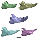
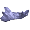
The present 3D Dataset contains the 3D models analyzed in 3D Finite Element Analysis and Geometric Morphometrics of Sloths (Xenarthra, Folivora) Mandibles Show Insights on the Dietary Specializations of Fossil Taxa. Journal of South American Earth Sciences. https://doi.org/10.1016/j.jsames.2023.104445
Mylodon darwinii CAV 379 View specimen
|
M3#1159Right hemimandible Type: "3D_surfaces"doi: 10.18563/m3.sf.1159 state:published |
Download 3D surface file |
Scelidotherium leptocephalum MNHN-M 137,722 View specimen
|
M3#1160Mandible Type: "3D_surfaces"doi: 10.18563/m3.sf.1160 state:published |
Download 3D surface file |
Glossotherium robustum MNHN-M 914 View specimen
|
M3#1161Mandible Type: "3D_surfaces"doi: 10.18563/m3.sf.1161 state:published |
Download 3D surface file |
Lestodon armatus MPAC 899 View specimen
|
M3#1162Mandible Type: "3D_surfaces"doi: 10.18563/m3.sf.1162 state:published |
Download 3D surface file |
Valgipes bucklandi NHMD.Z.M.K. 1/1845:3540 View specimen
|
M3#1163Mandible Type: "3D_surfaces"doi: 10.18563/m3.sf.1163 state:published |
Download 3D surface file |
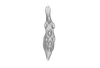
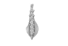
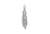
This contribution contains the 3D models of a set of Famennian conodont elements belonging to the species Icriodus alternatus analyzed in the following publication: Girard et al. 2022: Deciphering the morphological variation and its ontogenetic dynamics in the Late Devonian conodont Icriodus alternatus.
Icriodus alternatus UM BUS 031 View specimen
|
M3#887conodont element Type: "3D_surfaces"doi: 10.18563/m3.sf.887 state:published |
Download 3D surface file |
Icriodus alternatus UM BUS 032 View specimen
|
M3#888conodont element Type: "3D_surfaces"doi: 10.18563/m3.sf.888 state:published |
Download 3D surface file |
Icriodus alternatus UM BUS 033 View specimen
|
M3#889conodont element Type: "3D_surfaces"doi: 10.18563/m3.sf.889 state:published |
Download 3D surface file |
Icriodus alternatus UM BUS 034 View specimen
|
M3#890conodont element Type: "3D_surfaces"doi: 10.18563/m3.sf.890 state:published |
Download 3D surface file |
Icriodus alternatus UM BUS 035 View specimen
|
M3#891conodont element Type: "3D_surfaces"doi: 10.18563/m3.sf.891 state:published |
Download 3D surface file |
Icriodus alternatus UM BUS 036 View specimen
|
M3#892conodont element Type: "3D_surfaces"doi: 10.18563/m3.sf.892 state:published |
Download 3D surface file |
Icriodus alternatus UM BUS 037 View specimen
|
M3#893conodont element Type: "3D_surfaces"doi: 10.18563/m3.sf.893 state:published |
Download 3D surface file |
Icriodus alternatus UM BUS 038 View specimen
|
M3#894conodont element Type: "3D_surfaces"doi: 10.18563/m3.sf.894 state:published |
Download 3D surface file |
Icriodus alternatus UM BUS 039 View specimen
|
M3#895conodont element Type: "3D_surfaces"doi: 10.18563/m3.sf.895 state:published |
Download 3D surface file |
Icriodus alternatus UM BUS 040 View specimen
|
M3#896conodont element Type: "3D_surfaces"doi: 10.18563/m3.sf.896 state:published |
Download 3D surface file |
Icriodus alternatus UM BUS 041 View specimen
|
M3#897conodont element Type: "3D_surfaces"doi: 10.18563/m3.sf.897 state:published |
Download 3D surface file |
Icriodus alternatus UM BUS 042 View specimen
|
M3#898conodont element Type: "3D_surfaces"doi: 10.18563/m3.sf.898 state:published |
Download 3D surface file |
Icriodus alternatus UM BUS 043 View specimen
|
M3#899conodont element Type: "3D_surfaces"doi: 10.18563/m3.sf.899 state:published |
Download 3D surface file |
Icriodus alternatus UM BUS 044 View specimen
|
M3#900conodont element Type: "3D_surfaces"doi: 10.18563/m3.sf.900 state:published |
Download 3D surface file |
Icriodus alternatus UM BUS 045 View specimen
|
M3#901conodont element Type: "3D_surfaces"doi: 10.18563/m3.sf.901 state:published |
Download 3D surface file |
The present 3D Dataset contains the 3D model analyzed in Wazir, W. A., Sehgal, R. K., Čerňanský, A., Patnaik, R., Kumar, N., Singh, A. P. and Singh, N. P. 2022. A find from the Ladakh Himalaya reveals a survival of madtsoiid snakes (Serpentes, Madtsoiidae) in India through the late Oligocene. Journal of Vertebrate Paleontology, 41(6), e2058401. https://doi.org/10.1080/02724634.2021.2058401
indet. indet. WIMF/A 4816 View specimen

|
M3#1754Vertebra Type: "3D_surfaces"doi: 10.18563/m3.sf.1754 state:published |
Download 3D surface file |
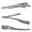
The present 3D dataset contains the 3D models of the holotype of Proterochampsa nodosa that were built and analysed in “Redescription, taxonomic revaluation, and phylogenetic affinities of Proterochampsa nodosa (Archosauriformes: Proterochampsidae), early Late Triassic of Candelaria Sequence (Santa Maria Supersequence)”.
Proterochampsa nodosa MCP 1694-PV View specimen
|
M3#9743D models of Proterochampsa nodosa. Model 1: skull. Model 2: mandible. Model 3: left mandibular ramus. Type: "3D_surfaces"doi: 10.18563/m3.sf.974 state:published |
Download 3D surface file |
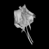
Our knowledge of the external brain morphology of the late Eocene artiodactyl ungulate Mixtotherium, relies on a plaster model realized on a specimen from the Victor Brun Museum in Montauban (France) and described by Dechaseaux (1973). Here, based on micro CT-scan data, we virtually reconstruct the 3D cast of the empty cavity of the partial cranium MA PHQ 716 from the Victor Brun Museum and compare it to the plaster model illustrated and described by Dechaseaux (1973). Indeed, the specimen from which the original plaster endocast originates was not identified by Dechaseaux by a specimen number. We confirm here that the studied specimen was indeed the one described and illustrated by Dechaseaux (1973). We also reconstruct a second, more detailed, model providing additional morphological and quantitative observations made available by micro CT scan investigation such as precisions on the neopallium folding and endocranial volumes.
Mixtotherium cuspidatum MA PHQ 716 View specimen
|
M3#857endocast of the brain cavity Type: "3D_surfaces"doi: 10.18563/m3.sf.857 state:published |
Download 3D surface file |
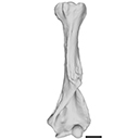
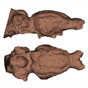
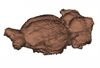
The present 3D Dataset contains the 3D model analyzed in the following publication: occurrence of the ground sloth Nothrotheriops (Xenarthra, Folivora) in the Late Pleistocene of Uruguay: New information on its dietary and habitat preferences based on stable isotope analysis. Journal of Mammalian Evolution. https://doi.org/10.1007/s10914-023-09660-w
Nothrotheriops sp. CAV 1466 View specimen
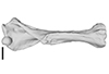
|
M3#1129Left humerus Type: "3D_surfaces"doi: 10.18563/m3.sf.1129 state:published |
Download 3D surface file |
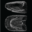
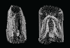
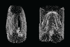
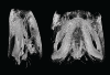
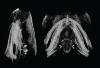
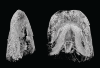
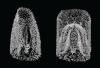
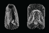
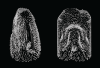
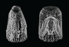
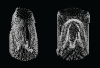
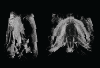
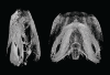
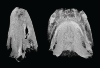
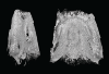
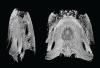
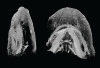
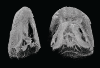
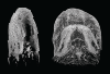
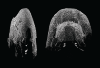
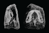
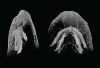
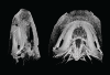
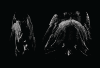
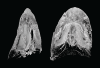
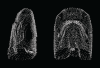
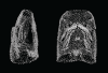
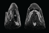
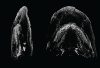
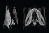
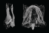
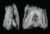
The present dataset contains the 3D models analyzed in Berio, F., Bayle, Y., Baum, D., Goudemand, N., and Debiais-Thibaud, M. 2022. Hide and seek shark teeth in Random Forests: Machine learning applied to Scyliorhinus canicula. It contains the head surfaces of 56 North Atlantic and Mediterranean small-spotted catsharks Scyliorhinus canicula, from which tooth surfaces were further extracted to perform geometric morphometrics and machine learning.
Scyliorhinus canicula 081118A View specimen
|
M3#941Head of a 10.6 cm long Scyliorhinus canicula female from a North Atlantic population. Type: "3D_surfaces"doi: 10.18563/m3.sf.941 state:published |
Download 3D surface file |
Scyliorhinus canicula 081118B View specimen
|
M3#942Head of a 11.0 cm long Scyliorhinus canicula female from a North Atlantic population. Type: "3D_surfaces"doi: 10.18563/m3.sf.942 state:published |
Download 3D surface file |
Scyliorhinus canicula 200118I View specimen
|
M3#959Head of a 45.0 cm long Scyliorhinus canicula female from a Mediterranean population. Type: "3D_surfaces"doi: 10.18563/m3.sf.959 state:published |
Download 3D surface file |
Scyliorhinus canicula 200118H View specimen
|
M3#958Head of a 47.0 cm long Scyliorhinus canicula female from a Mediterranean population. Type: "3D_surfaces"doi: 10.18563/m3.sf.958 state:published |
Download 3D surface file |
Scyliorhinus canicula 200118G View specimen
|
M3#957Head of a 40.0 cm long Scyliorhinus canicula female from a Mediterranean population. Type: "3D_surfaces"doi: 10.18563/m3.sf.957 state:published |
Download 3D surface file |
Scyliorhinus canicula 081118C View specimen
|
M3#940Head of a 11.2 cm long Scyliorhinus canicula female from a North Atlantic population. Type: "3D_surfaces"doi: 10.18563/m3.sf.940 state:published |
Download 3D surface file |
Scyliorhinus canicula 081118D View specimen
|
M3#939Head of a 10.2 cm long Scyliorhinus canicula female from a North Atlantic population. Type: "3D_surfaces"doi: 10.18563/m3.sf.939 state:published |
Download 3D surface file |
Scyliorhinus canicula 081118E View specimen
|
M3#938Head of a 12.0 cm long Scyliorhinus canicula male from a North Atlantic population. Type: "3D_surfaces"doi: 10.18563/m3.sf.938 state:published |
Download 3D surface file |
Scyliorhinus canicula 081118F View specimen
|
M3#937Head of a 10.7 cm long Scyliorhinus canicula male from a North Atlantic population. Type: "3D_surfaces"doi: 10.18563/m3.sf.937 state:published |
Download 3D surface file |
Scyliorhinus canicula 081118G View specimen
|
M3#936Head of a 10.8 cm long Scyliorhinus canicula male from a North Atlantic population. Type: "3D_surfaces"doi: 10.18563/m3.sf.936 state:published |
Download 3D surface file |
Scyliorhinus canicula 200118F View specimen
|
M3#935Head of a 41.5 cm long Scyliorhinus canicula female from a Mediterranean population. Type: "3D_surfaces"doi: 10.18563/m3.sf.935 state:published |
Download 3D surface file |
Scyliorhinus canicula 200118E View specimen
|
M3#934Head of a 40.0 cm long Scyliorhinus canicula female from a Mediterranean population. Type: "3D_surfaces"doi: 10.18563/m3.sf.934 state:published |
Download 3D surface file |
Scyliorhinus canicula 200118D View specimen
|
M3#933Head of a 42.0 cm long Scyliorhinus canicula male from a Mediterranean population. Type: "3D_surfaces"doi: 10.18563/m3.sf.933 state:published |
Download 3D surface file |
Scyliorhinus canicula 200118C View specimen
|
M3#943Head of a 41.0 cm long Scyliorhinus canicula male from a Mediterranean population. Type: "3D_surfaces"doi: 10.18563/m3.sf.943 state:published |
Download 3D surface file |
Scyliorhinus canicula 200118B View specimen
|
M3#945Head of a 44.0 cm long Scyliorhinus canicula male from a Mediterranean population. Type: "3D_surfaces"doi: 10.18563/m3.sf.945 state:published |
Download 3D surface file |
Scyliorhinus canicula 200118A View specimen
|
M3#944Head of a 46.0 cm long Scyliorhinus canicula male from a Mediterranean population. Type: "3D_surfaces"doi: 10.18563/m3.sf.944 state:published |
Download 3D surface file |
Scyliorhinus canicula 030418A View specimen
|
M3#956Head of a 13.9 cm long Scyliorhinus canicula female from a North Atlantic population. Type: "3D_surfaces"doi: 10.18563/m3.sf.956 state:published |
Download 3D surface file |
Scyliorhinus canicula 030418B View specimen
|
M3#955Head of a 13.6 cm long Scyliorhinus canicula female from a North Atlantic population. Type: "3D_surfaces"doi: 10.18563/m3.sf.955 state:published |
Download 3D surface file |
Scyliorhinus canicula 030418C View specimen
|
M3#954Head of a 13.4 cm long Scyliorhinus canicula male from a North Atlantic population. Type: "3D_surfaces"doi: 10.18563/m3.sf.954 state:published |
Download 3D surface file |
Scyliorhinus canicula 030418D View specimen
|
M3#953Head of a 13.2 cm long Scyliorhinus canicula male from a North Atlantic population. Type: "3D_surfaces"doi: 10.18563/m3.sf.953 state:published |
Download 3D surface file |
Scyliorhinus canicula 071118A View specimen
|
M3#952Head of a 36.0 cm long Scyliorhinus canicula female from a North Atlantic population. Type: "3D_surfaces"doi: 10.18563/m3.sf.952 state:published |
Download 3D surface file |
Scyliorhinus canicula 071118B View specimen
|
M3#951Head of a 33.0 cm long Scyliorhinus canicula female from a North Atlantic population. Type: "3D_surfaces"doi: 10.18563/m3.sf.951 state:published |
Download 3D surface file |
Scyliorhinus canicula 071118C View specimen
|
M3#950Head of a 32.0 cm long Scyliorhinus canicula female from a North Atlantic population. Type: "3D_surfaces"doi: 10.18563/m3.sf.950 state:published |
Download 3D surface file |
Scyliorhinus canicula 071118D View specimen
|
M3#949Head of a 35.0 cm long Scyliorhinus canicula male from a North Atlantic population. Type: "3D_surfaces"doi: 10.18563/m3.sf.949 state:published |
Download 3D surface file |
Scyliorhinus canicula 071118E View specimen
|
M3#948Head of a 35.0 cm long Scyliorhinus canicula male from a North Atlantic population. Type: "3D_surfaces"doi: 10.18563/m3.sf.948 state:published |
Download 3D surface file |
Scyliorhinus canicula 071118F View specimen
|
M3#947Head of a 33.0 cm long Scyliorhinus canicula male from a North Atlantic population. Type: "3D_surfaces"doi: 10.18563/m3.sf.947 state:published |
Download 3D surface file |
Scyliorhinus canicula 121118G View specimen
|
M3#946Head of a 36.0 cm long Scyliorhinus canicula female from a North Atlantic population. Type: "3D_surfaces"doi: 10.18563/m3.sf.946 state:published |
Download 3D surface file |
Scyliorhinus canicula 121118H View specimen
|
M3#932Head of a 35.0 cm long Scyliorhinus canicula female from a North Atlantic population. Type: "3D_surfaces"doi: 10.18563/m3.sf.932 state:published |
Download 3D surface file |
Scyliorhinus canicula 121118I View specimen
|
M3#931Head of a 33.0 cm long Scyliorhinus canicula male from a North Atlantic population. Type: "3D_surfaces"doi: 10.18563/m3.sf.931 state:published |
Download 3D surface file |
Scyliorhinus canicula 121118J View specimen
|
M3#917Head of a 36.0 cm long Scyliorhinus canicula male from a North Atlantic population. Type: "3D_surfaces"doi: 10.18563/m3.sf.917 state:published |
Download 3D surface file |
Scyliorhinus canicula 180118A View specimen
|
M3#916Head of a 57.0 cm long Scyliorhinus canicula female from a North Atlantic population. Type: "3D_surfaces"doi: 10.18563/m3.sf.916 state:published |
Download 3D surface file |
Scyliorhinus canicula 180118B View specimen
|
M3#915Head of a 58.0 cm long Scyliorhinus canicula female from a North Atlantic population. Type: "3D_surfaces"doi: 10.18563/m3.sf.915 state:published |
Download 3D surface file |
Scyliorhinus canicula 180118C View specimen
|
M3#911Head of a 58.5 cm long Scyliorhinus canicula female from a North Atlantic population. Type: "3D_surfaces"doi: 10.18563/m3.sf.911 state:published |
Download 3D surface file |
Scyliorhinus canicula 180118D View specimen
|
M3#914Head of a 56.0 cm long Scyliorhinus canicula male from a North Atlantic population. Type: "3D_surfaces"doi: 10.18563/m3.sf.914 state:published |
Download 3D surface file |
Scyliorhinus canicula 180118E View specimen
|
M3#913Head of a 58.0 cm long Scyliorhinus canicula male from a North Atlantic population. Type: "3D_surfaces"doi: 10.18563/m3.sf.913 state:published |
Download 3D surface file |
Scyliorhinus canicula 180118F View specimen
|
M3#912Head of a 59.0 cm long Scyliorhinus canicula male from a North Atlantic population. Type: "3D_surfaces"doi: 10.18563/m3.sf.912 state:published |
Download 3D surface file |
Scyliorhinus canicula 270918A View specimen
|
M3#910Head of a 56.0 cm long Scyliorhinus canicula male from a North Atlantic population. Type: "3D_surfaces"doi: 10.18563/m3.sf.910 state:published |
Download 3D surface file |
Scyliorhinus canicula 270918B View specimen
|
M3#908Head of a 59.5 cm long Scyliorhinus canicula male from a North Atlantic population. Type: "3D_surfaces"doi: 10.18563/m3.sf.908 state:published |
Download 3D surface file |
Scyliorhinus canicula 270918C View specimen
|
M3#909Head of a 63.0 cm long Scyliorhinus canicula female from a North Atlantic population. Type: "3D_surfaces"doi: 10.18563/m3.sf.909 state:published |
Download 3D surface file |
Scyliorhinus canicula 270918D View specimen
|
M3#907Head of a 64.0 cm long Scyliorhinus canicula female from a North Atlantic population. Type: "3D_surfaces"doi: 10.18563/m3.sf.907 state:published |
Download 3D surface file |
Scyliorhinus canicula 12111931 View specimen
|
M3#905Head of a 9.5 cm long Scyliorhinus canicula male from a Mediterranean population. Type: "3D_surfaces"doi: 10.18563/m3.sf.905 state:published |
Download 3D surface file |
Scyliorhinus canicula 12111933 View specimen
|
M3#906Head of a 9.5 cm long Scyliorhinus canicula female from a Mediterranean population. Type: "3D_surfaces"doi: 10.18563/m3.sf.906 state:published |
Download 3D surface file |
Scyliorhinus canicula 190118A View specimen
|
M3#918Head of a 8.8 cm long Scyliorhinus canicula female from a Mediterranean population. Type: "3D_surfaces"doi: 10.18563/m3.sf.918 state:published |
Download 3D surface file |
Scyliorhinus canicula 190118C View specimen
|
M3#930Head of a 9.0 cm long Scyliorhinus canicula female from a Mediterranean population. Type: "3D_surfaces"doi: 10.18563/m3.sf.930 state:published |
Download 3D surface file |
Scyliorhinus canicula 190118D View specimen
|
M3#929Head of a 8.9 cm long Scyliorhinus canicula male from a Mediterranean population. Type: "3D_surfaces"doi: 10.18563/m3.sf.929 state:published |
Download 3D surface file |
Scyliorhinus canicula 190118F View specimen
|
M3#928Head of a 9.1 cm long Scyliorhinus canicula male from a Mediterranean population. Type: "3D_surfaces"doi: 10.18563/m3.sf.928 state:published |
Download 3D surface file |
Scyliorhinus canicula 060718A View specimen
|
M3#927Head of a 25.5 cm long Scyliorhinus canicula male from a Mediterranean population. Type: "3D_surfaces"doi: 10.18563/m3.sf.927 state:published |
Download 3D surface file |
Scyliorhinus canicula 060718B View specimen
|
M3#926Head of a 23.0 cm long Scyliorhinus canicula female from a Mediterranean population. Type: "3D_surfaces"doi: 10.18563/m3.sf.926 state:published |
Download 3D surface file |
Scyliorhinus canicula 060718C View specimen
|
M3#925Head of a 28.0 cm long Scyliorhinus canicula male from a Mediterranean population. Type: "3D_surfaces"doi: 10.18563/m3.sf.925 state:published |
Download 3D surface file |
Scyliorhinus canicula 060718D View specimen
|
M3#924Head of a 21.0 cm long Scyliorhinus canicula male from a Mediterranean population. Type: "3D_surfaces"doi: 10.18563/m3.sf.924 state:published |
Download 3D surface file |
Scyliorhinus canicula 060718E View specimen

|
M3#923Head of a 23.5 cm long Scyliorhinus canicula male from a Mediterranean population. Type: "3D_surfaces"doi: 10.18563/m3.sf.923 state:published |
Download 3D surface file |
Scyliorhinus canicula 060718F View specimen
|
M3#922Head of a 22.5 cm long Scyliorhinus canicula female from a Mediterranean population. Type: "3D_surfaces"doi: 10.18563/m3.sf.922 state:published |
Download 3D surface file |
Scyliorhinus canicula 121218A View specimen
|
M3#921Head of a 31.0 cm long Scyliorhinus canicula female from a Mediterranean population. Type: "3D_surfaces"doi: 10.18563/m3.sf.921 state:published |
Download 3D surface file |
Scyliorhinus canicula 121218B View specimen
|
M3#920Head of a 31.0 cm long Scyliorhinus canicula female from a Mediterranean population. Type: "3D_surfaces"doi: 10.18563/m3.sf.920 state:published |
Download 3D surface file |
Scyliorhinus canicula 121218C View specimen
|
M3#919Head of a 31.0 cm long Scyliorhinus canicula female from a Mediterranean population. Type: "3D_surfaces"doi: 10.18563/m3.sf.919 state:published |
Download 3D surface file |
Scyliorhinus canicula 121218D View specimen
|
M3#904Head of a 31.0 cm long Scyliorhinus canicula male from a Mediterranean population. Type: "3D_surfaces"doi: 10.18563/m3.sf.904 state:published |
Download 3D surface file |
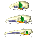
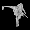
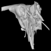
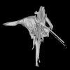
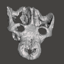
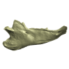
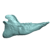
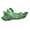
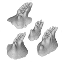
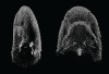
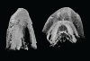
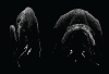
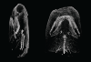
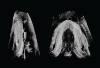
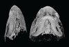
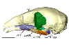
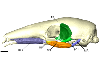
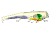
The present 3D Dataset contains the 3D models described in “Comparative masticatory myology in anteaters and its implications for interpreting morphological convergence in myrmecophagous placentals”.
Cyclopes didactylus M1571_JAG View specimen
|
M3#522Skull, mandible, and muscles of Cyclopes didactylus Type: "3D_surfaces"doi: 10.18563/m3.sf.522 state:published |
Download 3D surface file |
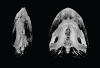
Tamandua tetradactyla M3075_JAG View specimen
|
M3#524Skull, left mandibles, and muscles of Tamandua tetradactyla. Type: "3D_surfaces"doi: 10.18563/m3.sf.524 state:published |
Download 3D surface file |
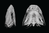
Myrmecophaga tridactyla M3023_JAG View specimen
|
M3#523Skull, left mandible and muscles of Myrmecophaga tridactyla. Type: "3D_surfaces"doi: 10.18563/m3.sf.523 state:published |
Download 3D surface file |


