Explodable 3D Dog Skull for Veterinary Education
3D models of Ocnotherium skull
3D models of Kalakocetus, the earliest Cetacea
3D GM dataset of bird skeletal variation
Skeletal embryonic development in the catshark
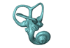
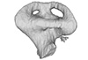
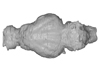
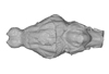
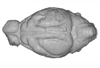
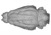
Bony connexions of the petrosal bone of extant hippos
bony labyrinth (14) , inner ear (11) , geometric morphometrics (10) , CT-scan (10) , Eocene (10) , Micro-CT (9) , Miocene (8)
Lionel Hautier (24) , Maëva Judith Orliac (23) , Laurent Marivaux (18) , Renaud Lebrun (14) , Rodolphe Tabuce (14) , Pierre-Olivier Antoine (13) , Bastien Mennecart (13)
|
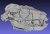
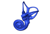
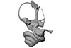
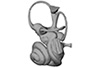
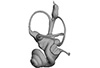
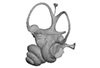
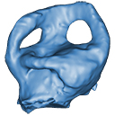
3D model related to the publication: The inner ear of Megatherium and the evolution of the vestibular system in sloths.Guillaume Billet
Published online: 24/02/2015 |

|
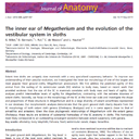
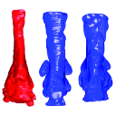
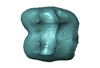
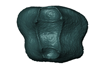
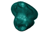
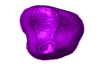
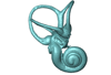
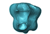
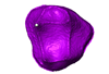
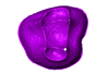
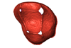
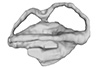
M3#14This model corresponds to a virtually reconstructed bony labyrinth of the right inner ear of the skull MNHN-F-PAM 276, attributed to the extinct giant ground sloth Megatherium americanum. The fossil comes from Pleistocene deposits at Rio Salado (Prov. Buenos Aires, Argentina). The bony labyrinth of Megatherium shows semicircular canals that are proportionally much larger than in the modern two-toed and three-toed sloths. The cochlea in Megatherium shows 2.5 turns, which is a rather high value within Xenarthra. Overall, the shape of the bony labyrinth of Megatherium resembles more that of extant armadillos than that of its extant sloth relatives. Type: "3D_surfaces"doi: 10.18563/m3.sf14 state:published |
Download 3D surface file |
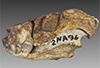
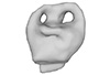
This contribution contains the 3D model of an endocranial cast analyzed in “A 10 ka intentionally deformed human skull from Northeast Asia”. There are many studies on the morphological characteristics of intentional cranial deformation (ICD), but few related 3D models were published. Here, we present the surface model of an intentionally deformed 10 ka human cranium for further research on ICD practice. The 3D model of the endocranial cast of this ICD cranium was discovered near Harbin City, Province Heilongjiang, Northeast China. The fossil preserved only the frontal, parietal, and occipital bones. To complete the endocast model of the specimen, we printed a 3D model and used modeling clay to reconstruct the missing part based on the general form of the modern human endocast morphology.
Homo sapiens IVPP-PA1616 View specimen
|
M3#972The frontal region of the endocast is flattened, probably formed by the constant pressure on the frontal bone during growth. There is a well-developed frontal crest on the endocranial surface. The endocast widens posteriorly from the frontal lobe. The widest point of the endocast is at the lateral border of the parietal lobe. The lower parietal areas display a marked lateral expansion. The overall shape of the endocast is asymmetrical, with the left side of the parietal lobe being more laterally expanded than the right side. Like the frontal lobe, the occipital lobe is also anteroposteriorly flattened. Type: "3D_surfaces"doi: 10.18563/m3.sf.972 state:published |
Download 3D surface file |
|
M3#976The original endocranial cast model (with texture) of IVPP-PA1616. It shows the original structures of the specimen, and was not altered in any way. Type: "3D_surfaces"doi: 10.18563/m3.sf.976 state:published |
Download 3D surface file |
The present 3D Dataset contains the 3D models of brain endocast of traversodontid cynodonts studied in: Pavanatto et al. 2019. Virtual reconstruction of cranial endocasts of traversodontid cynodonts (Eucynodontia: Gomphodontia) from the upper Triassic of Southern Brazil. Journal of Morphology. https://doi.org/10.1002/jmor.21029
Siriusgnathus niemeyerorum CAPPA/UFSM 0032 View specimen
|
M3#4253D model of the brain endocast Type: "3D_surfaces"doi: 10.18563/m3.sf.425 state:published |
Download 3D surface file |
Exaeretodon riograndensis CAPPA/UFSM 0030 View specimen

|
M3#4263D model of the brain endocast Type: "3D_surfaces"doi: 10.18563/m3.sf.426 state:published |
Download 3D surface file |
Exaeretodon riograndensis CAPPA/UFSM 0227 View specimen
|
M3#4273D model of the brain endocast Type: "3D_surfaces"doi: 10.18563/m3.sf.427 state:published |
Download 3D surface file |
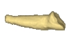
This note presents the 3D model of the hemi-mandible UM-PAT 159 of the MP7 Diacodexis species D. cf. gigasei and 3D models corresponding to the restoration of the ascending ramus, broken on the original specimen, and to a restoration of a complete mandible based on the preserved left hemi-mandible.
Diacodexis cf. gigasei UMPAT159 View specimen
|
M3#3153D models of UM PAT 159 after the restoration of the ascending ramus Type: "3D_surfaces"doi: 10.18563/m3.sf.315 state:published |
Download 3D surface file |
|
M3#316restoration of a complete mandible based on the preserved left hemi-mandible UM PAT 159 Type: "3D_surfaces"doi: 10.18563/m3.sf.316 state:published |
Download 3D surface file |
|
M3#3173D model of the hemi-mandible UM PAT 159 Type: "3D_surfaces"doi: 10.18563/m3.sf.317 state:published |
Download 3D surface file |
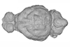
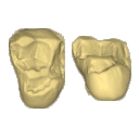
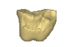
This contribution contains the 3D models of the isolated teeth attributed to stem representatives of the Cebuella and Cebus lineages (Cebuella sp. and Cebus sp.), described and figured in the following publication: Marivaux et al. (2016), Dental remains of cebid platyrrhines from the earliest late Miocene of Western Amazonia, Peru: macroevolutionary implications on the extant capuchin and marmoset lineages. American Journal of Physical Anthropology. http://dx.doi.org/10.1002/ajpa.23052
Cebus sp. MUSM-3243 View specimen
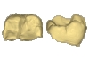
|
M3#2823D model of left lower m1 (lingual part) Type: "3D_surfaces"doi: 10.18563/m3.sf.282 state:published |
Download 3D surface file |
Cebuella sp. MUSM-3239 View specimen
|
M3#2833D model of left lower p4 Type: "3D_surfaces"doi: 10.18563/m3.sf.283 state:published |
Download 3D surface file |
Cebuella sp. MUSM-3240 View specimen
|
M3#2943D model of right upper P3 or P4 (buccal part) Type: "3D_surfaces"doi: 10.18563/m3.sf.294 state:published |
Download 3D surface file |
Cebuella sp. MUSM-3241 View specimen
|
M3#2953D model of right upper P2 Type: "3D_surfaces"doi: 10.18563/m3.sf.295 state:published |
Download 3D surface file |
Cebuella sp. MUSM-3242 View specimen
|
M3#2963D model of upper I2 Type: "3D_surfaces"doi: 10.18563/m3.sf.296 state:published |
Download 3D surface file |
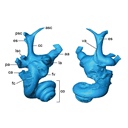
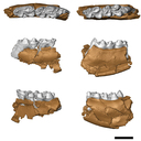
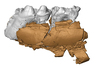
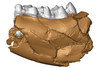
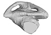
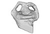
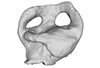
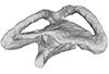
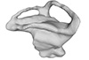
The present 3D Dataset contains the 3D models analyzed in Costeur L., Mennecart B., Müller B., Schulz G., 2016. Prenatal growth stages show the development of the ruminant bony labyrinth and petrosal bone. Journal of Anatomy. https://doi.org/10.1111/joa.12549
Bos taurus NMB3038 View specimen
|
M3#124Right bony labyrinth of a Bos taurus foetus (gestational age 115 days) Type: "3D_surfaces"doi: 10.18563/m3.sf.124 state:published |
Download 3D surface file |
Bos taurus NMB3367 View specimen
|
M3#125Right bony labyrinth of a Bos taurus foetus (gestational age 165 days) Type: "3D_surfaces"doi: 10.18563/m3.sf.125 state:published |
Download 3D surface file |
Bos taurus NMB3365 View specimen
|
M3#126Right bony labyrinth of a Bos taurus foetus (gestational age 210 days) Type: "3D_surfaces"doi: 10.18563/m3.sf.126 state:published |
Download 3D surface file |
Bos taurus NMB2855 View specimen
|
M3#127Right bony labyrinth of a Bos taurus foetus (gestational age 260 days) Type: "3D_surfaces"doi: 10.18563/m3.sf.127 state:published |
Download 3D surface file |
Bos taurus NMB1037 View specimen
|
M3#128Left bony labyrinth of an adult Bos taurus Type: "3D_surfaces"doi: 10.18563/m3.sf.128 state:published |
Download 3D surface file |
The present 3D Dataset contains the 3D models analyzed in: Hirose, A., Nakashima, T., Yamada, S., Uwabe, C., Kose, K., Takakuwa, T. 2012. Embryonic liver morphology and morphometry by magnetic resonance microscopic imaging. Anat Rec (Hoboken) 295, 51-59. doi: 10.1002/ar.21496
Homo sapiens KC-CS14LIV1387 View specimen
|
M3#64Human liver at Carnegie Stage (CS) 14 Type: "3D_surfaces"doi: 10.18563/m3.sf.64 state:published |
Download 3D surface file |
Homo sapiens KC-CS15LIV5074 View specimen
|
M3#65Human liver at Carnegie Stage (CS) 15 Type: "3D_surfaces"doi: 10.18563/m3.sf.65 state:published |
Download 3D surface file |
Homo sapiens KC-CS16LIV2578 View specimen
|
M3#66Human liver at Carnegie Stage (CS) 16 Type: "3D_surfaces"doi: 10.18563/m3.sf.66 state:published |
Download 3D surface file |
Homo sapiens KC-CS17LIV17832 View specimen
|
M3#67Human liver at Carnegie Stage (CS) 17 Type: "3D_surfaces"doi: 10.18563/m3.sf.67 state:published |
Download 3D surface file |
Homo sapiens KC-CS18LIV21124 View specimen
|
M3#68Human liver at Carnegie Stage (CS) 18 Type: "3D_surfaces"doi: 10.18563/m3.sf.68 state:published |
Download 3D surface file |
Homo sapiens KC-CS19LIV14353 View specimen

|
M3#69Human liver at Carnegie Stage (CS) 19 Type: "3D_surfaces"doi: 10.18563/m3.sf.69 state:published |
Download 3D surface file |
Homo sapiens KC-CS20LIV20701 View specimen
|
M3#70Human liver at Carnegie Stage (CS) 20 Type: "3D_surfaces"doi: 10.18563/m3.sf.70 state:published |
Download 3D surface file |
Homo sapiens KC-CS21LIV25858 View specimen
|
M3#71Human liver at Carnegie Stage (CS) 21 Type: "3D_surfaces"doi: 10.18563/m3.sf.71 state:published |
Download 3D surface file |
Homo sapiens KC-CS22LIV22226 View specimen
|
M3#72Human liver at Carnegie Stage (CS) 22 Type: "3D_surfaces"doi: 10.18563/m3.sf.72 state:published |
Download 3D surface file |
Homo sapiens KC-CS23LIV25704 View specimen
|
M3#73Human liver at Carnegie Stage (CS) 23 Type: "3D_surfaces"doi: 10.18563/m3.sf.73 state:published |
Download 3D surface file |
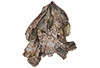
This contribution contains 3D models of the holotype of a new species of long-nosed armadillos, the Guianan long-nosed armadillo (Dasypus guianensis) described in the following publication: Barthe M., Rancilhac L., Arteaga M. C., Feijó A., Tilak M.-K., Justy F., Loughry W. J., McDonough C. M., de Thoisy B., Catzeflis F., Billet G., Hautier L., Nabholz B., and Delsuc F. 2024. Exon capture museomics deciphers the nine-banded armadillo species complex and identifies a new species endemic to the Guiana Shield. Systematic Biology, syae027. https://doi.org/10.1093/sysbio/syae027
Dasypus guianensis MNHN-ZM-MO-2001-1317 View specimen
|
M3#1200Skeleton and carapace Type: "3D_surfaces"doi: 10.18563/m3.sf.1200 state:published |
Download 3D surface file |
|
M3#1201Frontal sinuses Type: "3D_surfaces"doi: 10.18563/m3.sf.1201 state:published |
Download 3D surface file |
The present 3D Dataset contains the 3D models analyzed in Mennecart B., Wazir W.A., Sehgal R.K., Patnaik R., Singh N.P., Kumar N, and Nanda A.C. 2021. New remains of Nalamaeryx (Tragulidae, Mammalia) from the Ladakh Himalaya and their phylogenetical and palaeoenvironmental implications. Historical Biology. https://doi.org/10.1080/08912963.2021.2014479
Nalameryx savagei WIMF/A4801 View specimen
|
M3#766Nalameryx savagei, Partial lower right jaw preserving m2 and m3. Type: "3D_surfaces"doi: 10.18563/m3.sf.766 state:published |
Download 3D surface file |
Nalameryx savagei WIMF/A4802 View specimen
|
M3#767Nalameryx savagei, partial lower right jaw preserving m2 and m3 Type: "3D_surfaces"doi: 10.18563/m3.sf.767 state:published |
Download 3D surface file |
The present 3D Dataset contains the 3D models analyzed in Hendrickx, C., Gaetano, L. C., Choiniere, J., Mocke, H. and Abdala, F. in press. A new traversodontid cynodont with a peculiar postcanine dentition from the Middle/Late Triassic of Namibia and dental evolution in basal gomphodonts. Journal of Systematic Palaeontology.
Etjoia dentitransitus GSN F1591 View specimen
|
M3#557Surface model derived from µCT data of the holotype of Etjoia dentitransitus Type: "3D_surfaces"doi: 10.18563/m3.sf.557 state:published |
Download 3D surface file |
|
M3#558Photogrammetric 3D surface model of the postcanines of the Holotype of Etjoia dentitransitus Type: "3D_surfaces"doi: 10.18563/m3.sf.558 state:published |
Download 3D surface file |
|
M3#559Photogrammetric 3D surface model of the Holotype of Etjoia dentitransitus Type: "3D_surfaces"doi: 10.18563/m3.sf.559 state:published |
Download 3D surface file |
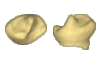
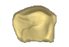
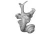
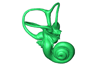
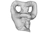
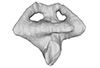
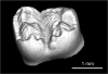
The present 3D Dataset contains the 3D models of the enamel-dentine junctions of upper third molars and of the bony labyrinths of the extant cercopithecoid specimens analyzed in the following publication: Beaudet, A., Dumoncel, J., Thackeray, J.F., Bruxelles, L., Duployer, B., Tenailleau, C., Bam, L., Hoffman, J., de Beer, F., Braga, J.: Upper third molar internal structural organization and semicircular canal morphology in Plio-Pleistocene South African cercopithecoids. Journal of Human Evolution 95, 104-120. https://doi.org/10.1016/j.jhevol.2016.04.004
Cercocebus atys 81.007-M-0041 View specimen
|
M3#4453D model of the enamel-dentine junction of the right upper third molar. Type: "3D_surfaces"doi: 10.18563/m3.sf.445 state:published |
Download 3D surface file |
Cercocebus torquatus 73.018-M-0359 View specimen
|
M3#4463D model of the enamel-dentine junction of the right upper third molar. Type: "3D_surfaces"doi: 10.18563/m3.sf.446 state:published |
Download 3D surface file |

|
M3#4963D model of the left bony labyrinth. Type: "3D_surfaces"doi: 10.18563/m3.sf.496 state:published |
Download 3D surface file |
Mandrillus leucophaeus 73.029-M-0106 View specimen
|
M3#4473D model of the enamel-dentine junction of the right upper third molar. Type: "3D_surfaces"doi: 10.18563/m3.sf.447 state:published |
Download 3D surface file |

|
M3#4703D model of the right bony labyrinth. Type: "3D_surfaces"doi: 10.18563/m3.sf.470 state:published |
Download 3D surface file |
Lophocebus albigena 73.029-M-0109 View specimen
|
M3#4483D model of the enamel-dentine junction of the right upper third molar. Type: "3D_surfaces"doi: 10.18563/m3.sf.448 state:published |
Download 3D surface file |
|
M3#4713D model of the right bony labyrinth. Type: "3D_surfaces"doi: 10.18563/m3.sf.471 state:published |
Download 3D surface file |
Piliocolobus foai 91.060-M-0071 View specimen
|
M3#4493D model of the enamel-dentine junction of the right upper third molar. Type: "3D_surfaces"doi: 10.18563/m3.sf.449 state:published |
Download 3D surface file |

|
M3#4723D model of the right bony labyrinth. Type: "3D_surfaces"doi: 10.18563/m3.sf.472 state:published |
Download 3D surface file |
Colobus guereza 1215 View specimen
|
M3#4503D model of the enamel-dentine junction of the right upper third molar. Type: "3D_surfaces"doi: 10.18563/m3.sf.450 state:published |
Download 3D surface file |

|
M3#4733D model of the right bony labyrinth. Type: "3D_surfaces"doi: 10.18563/m3.sf.473 state:published |
Download 3D surface file |
Colobus guereza 2800 View specimen
|
M3#4513D model of the enamel-dentine junction of the right upper third molar. Type: "3D_surfaces"doi: 10.18563/m3.sf.451 state:published |
Download 3D surface file |

|
M3#4743D model of the right bony labyrinth. Type: "3D_surfaces"doi: 10.18563/m3.sf.474 state:published |
Download 3D surface file |
Papio cynocephalus kindae 3503 View specimen
|
M3#4523D model of the enamel-dentine junction of the right upper third molar. Type: "3D_surfaces"doi: 10.18563/m3.sf.452 state:published |
Download 3D surface file |

|
M3#4753D model of the right bony labyrinth. Type: "3D_surfaces"doi: 10.18563/m3.sf.475 state:published |
Download 3D surface file |
Erythrocebus patas 8452 View specimen
|
M3#4533D model of the enamel-dentine junction of the right upper third molar. Type: "3D_surfaces"doi: 10.18563/m3.sf.453 state:published |
Download 3D surface file |

|
M3#4763D model of the right bony labyrinth. Type: "3D_surfaces"doi: 10.18563/m3.sf.476 state:published |
Download 3D surface file |
Papio cynocephalus kindae 17979 View specimen
|
M3#4543D model of the enamel-dentine junction of the right upper third molar. Type: "3D_surfaces"doi: 10.18563/m3.sf.454 state:published |
Download 3D surface file |

|
M3#4773D model of the right bony labyrinth. Type: "3D_surfaces"doi: 10.18563/m3.sf.477 state:published |
Download 3D surface file |
Colobus angolensis 25456 View specimen
|
M3#4553D model of the enamel-dentine junction of the right upper third molar. Type: "3D_surfaces"doi: 10.18563/m3.sf.455 state:published |
Download 3D surface file |

|
M3#4783D model of the right bony labyrinth. Type: "3D_surfaces"doi: 10.18563/m3.sf.478 state:published |
Download 3D surface file |
Chlorocebus pygerythrus 37477 View specimen
|
M3#4563D model of the enamel-dentine junction of the right upper third molar. Type: "3D_surfaces"doi: 10.18563/m3.sf.456 state:published |
Download 3D surface file |
|
M3#4813D model of the right bony labyrinth. Type: "3D_surfaces"doi: 10.18563/m3.sf.481 state:published |
Download 3D surface file |
Chlorocebus pygerythrus 37478 View specimen
|
M3#4573D model of the enamel-dentine junction of the right upper third molar. Type: "3D_surfaces"doi: 10.18563/m3.sf.457 state:published |
Download 3D surface file |

|
M3#4823D model of the right bony labyrinth. Type: "3D_surfaces"doi: 10.18563/m3.sf.482 state:published |
Download 3D surface file |
Lophocebus albigena 37572 View specimen
|
M3#4583D model of the enamel-dentine junction of the right upper third molar. Type: "3D_surfaces"doi: 10.18563/m3.sf.458 state:published |
Download 3D surface file |

|
M3#4833D model of the right bony labyrinth. Type: "3D_surfaces"doi: 10.18563/m3.sf.483 state:published |
Download 3D surface file |
Lophocebus albigena 37579 View specimen
|
M3#4593D model of the enamel-dentine junction of the right upper third molar. Type: "3D_surfaces"doi: 10.18563/m3.sf.459 state:published |
Download 3D surface file |
Erythrocebus patas OST.2002-26 View specimen
|
M3#4603D model of the enamel-dentine junction of the right upper third molar. Type: "3D_surfaces"doi: 10.18563/m3.sf.460 state:published |
Download 3D surface file |
|
M3#4843D model of the right bony labyrinth. Type: "3D_surfaces"doi: 10.18563/m3.sf.484 state:published |
Download 3D surface file |
Mandrillus sphinx OST.AC.488 View specimen
|
M3#4613D model of the enamel-dentine junction of the right upper third molar. Type: "3D_surfaces"doi: 10.18563/m3.sf.461 state:published |
Download 3D surface file |
|
M3#4853D model of the left bony labyrinth. Type: "3D_surfaces"doi: 10.18563/m3.sf.485 state:published |
Download 3D surface file |
Macaca mulatta OST.AC.492 View specimen
|
M3#4623D model of the enamel-dentine junction of the right upper third molar. Type: "3D_surfaces"doi: 10.18563/m3.sf.462 state:published |
Download 3D surface file |
|
M3#4863D model of the right bony labyrinth. Type: "3D_surfaces"doi: 10.18563/m3.sf.486 state:published |
Download 3D surface file |
Chlorocebus aethiops OST.AC.523 View specimen
|
M3#4633D model of the enamel-dentine junction of the right upper third molar. Type: "3D_surfaces"doi: 10.18563/m3.sf.463 state:published |
Download 3D surface file |

|
M3#4913D model of the right bony labyrinth. Type: "3D_surfaces"doi: 10.18563/m3.sf.491 state:published |
Download 3D surface file |
Cercopithecus cephus OST.AC.533 View specimen
|
M3#4643D model of the enamel-dentine junction of the right upper third molar. Type: "3D_surfaces"doi: 10.18563/m3.sf.464 state:published |
Download 3D surface file |
|
M3#4933D model of the right bony labyrinth. Type: "3D_surfaces"doi: 10.18563/m3.sf.493 state:published |
Download 3D surface file |
Chlorocebus aethiops OST.AC.540 View specimen
|
M3#4653D model of the enamel-dentine junction of the right upper third molar. Type: "3D_surfaces"doi: 10.18563/m3.sf.465 state:published |
Download 3D surface file |

|
M3#4943D model of the right bony labyrinth. Type: "3D_surfaces"doi: 10.18563/m3.sf.494 state:published |
Download 3D surface file |
Mandrillus sphinx OST.AC.543 View specimen
|
M3#4663D model of the enamel-dentine junction of the right upper third molar. Type: "3D_surfaces"doi: 10.18563/m3.sf.466 state:published |
Download 3D surface file |
|
M3#4953D model of the right bony labyrinth. Type: "3D_surfaces"doi: 10.18563/m3.sf.495 state:published |
Download 3D surface file |
Cercocebus torquatus 73.018-M-389 View specimen

|
M3#4683D model of the right bony labyrinth. Type: "3D_surfaces"doi: 10.18563/m3.sf.468 state:published |
Download 3D surface file |
Mandrillus leucophaeus 73.029-M-0105 View specimen
|
M3#4693D model of the right bony labyrinth. Type: "3D_surfaces"doi: 10.18563/m3.sf.469 state:published |
Download 3D surface file |
Mandrillus leucophaeus 28425 View specimen

|
M3#4793D model of the right bony labyrinth. Type: "3D_surfaces"doi: 10.18563/m3.sf.479 state:published |
Download 3D surface file |
Cercocebus atys 28998 View specimen

|
M3#4803D model of the right bony labyrinth. Type: "3D_surfaces"doi: 10.18563/m3.sf.480 state:published |
Download 3D surface file |
Macaca sylvanus OST.AC.493 View specimen

|
M3#4873D model of the right bony labyrinth. Type: "3D_surfaces"doi: 10.18563/m3.sf.487 state:published |
Download 3D surface file |
Chlorocebus aethiops OST.AC.508 View specimen

|
M3#4883D model of the left bony labyrinth. Type: "3D_surfaces"doi: 10.18563/m3.sf.488 state:published |
Download 3D surface file |
Cercopithecus cephus OST.AC.515 View specimen

|
M3#4893D model of the right bony labyrinth. Type: "3D_surfaces"doi: 10.18563/m3.sf.489 state:published |
Download 3D surface file |
Colobus guereza OST.AC.519 View specimen

|
M3#4903D model of the right bony labyrinth. Type: "3D_surfaces"doi: 10.18563/m3.sf.490 state:published |
Download 3D surface file |
Macaca sp. OST.AC.532 View specimen

|
M3#4923D model of the left bony labyrinth. Type: "3D_surfaces"doi: 10.18563/m3.sf.492 state:published |
Download 3D surface file |
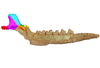
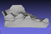
The present 3D Dataset contains the 3D models analyzed in: "a giant dapediid from the Late Triassic of Switzerland and insights into neopterygian phylogeny", Royal Society Open Science, https://doi.org/10.1098/rsos.180497
Scopulipiscis saxciput PIMUZ A/I 3026 View specimen

|
M3#1773D surfaces of the skull and endocranial spaces inside neurocranium, including the aortic canal, braincase, fossa bridgei, lateral cranial canal, nerves and other passageways, notochord, posterior myodome, and right semicircular canals. Type: "3D_surfaces"doi: 10.18563/m3.sf.177 state:published |
Download 3D surface file |

|
M3#178Scan of the neurocranium of PIMUZ A/I 3026 Type: "3D_CT"doi: 10.18563/m3.sf.178 state:published |
Download CT data |
The present 3D Dataset contains the 3D models analyzed in the article Mennecart, B., and L. Costeur. 2016. A Dorcatherium (Mammalia, Ruminantia, Middle Miocene) petrosal bone and the tragulid ear region. Journal of Vertebrate Paleontology 36(6), 1211665(1)-1211665(7). DOI: 10.1080/02724634.2016.1211665.
Tragulus javanicus 10028 View specimen
|
M3#1193D surface of the left bony labyrinth of Tragulus javanicus NMB 10028 Type: "3D_surfaces"doi: 10.18563/m3.sf.119 state:published |
Download 3D surface file |
Moschiola meminna C.2453 View specimen
|
M3#1203D surface of the left bony labyrinth of Moschiola meminna NMB C.2453 Type: "3D_surfaces"doi: 10.18563/m3.sf.120 state:published |
Download 3D surface file |
Hyemoschus aquaticus C.1930 View specimen
|
M3#1223D surface of the right bony labyrinth of Hyemoschus aquaticus NMB C.1930 Type: "3D_surfaces"doi: 10.18563/m3.sf.122 state:published |
Download 3D surface file |
Dorcatherium crassum San.15053 View specimen
|
M3#1233D surface of the right bony labyrinth of Dorcatherium crassum NMB San.15053 Type: "3D_surfaces"doi: 10.18563/m3.sf.123 state:published |
Download 3D surface file |
The present 3D Dataset contains the 3D models analyzed in "Neenan, J. M., Reich, T., Evers, S., Druckenmiller, P. S., Voeten, D. F. A. E., Choiniere, J. N., Barrett, P. M., Pierce, S. E. and Benson, R. B. J. Evolution of the sauropterygian labyrinth with increasingly pelagic lifestyles. Current Biology, 27." https://doi.org/10.1016/j.cub.2017.10.069
Amblyrhynchus cristatus OUMNH 11616 View specimen
|
M3#322Right labyrinth of Amblyrhynchus cristatus (OUMNH 11616). Type: "3D_surfaces"doi: 10.18563/m3.sf.322 state:published |
Download 3D surface file |
Augustasaurus hagdorni FMNH PR 1974 View specimen
|
M3#333Right labyrinth model of Augustasaurus FMNH PR 1974 Type: "3D_surfaces"doi: 10.18563/m3.sf.333 state:published |
Download 3D surface file |
Callawayasaurus colombiensis UCMP V-38349 / UCMP V-125328 View specimen
|
M3#331Composite left labyrinth of Callawayasaurus. The majority of the model is from the holotype (UCMP V-38349), but the anterior portion is formed from the right labyrinth (reflected) from the paratype (UCMP V-125328). Type: "3D_surfaces"doi: 10.18563/m3.sf.331 state:published |
Download 3D surface file |
Lepidochelys olivacea SMNS 11070 View specimen
|
M3#330Left labyrinth model of Lepidochelys SMNS 11070 Type: "3D_surfaces"doi: 10.18563/m3.sf.330 state:published |
Download 3D surface file |
Macrochelys temminckii FMNH 22111 View specimen
|
M3#334Left labyrinth model of Macrochelys FMNH 22111 Type: "3D_surfaces"doi: 10.18563/m3.sf.334 state:published |
Download 3D surface file |
Macroplata tenuiceps NHMUK R 5488 View specimen
|
M3#328Left labyrinth of Macroplata NHMUK R 5488 Type: "3D_surfaces"doi: 10.18563/m3.sf.328 state:published |
Download 3D surface file |
Microcleidus homalospondylus NHMUK 36184 View specimen
|
M3#327Right labyrinth model of Microcleidus NHMUK 36184 Type: "3D_surfaces"doi: 10.18563/m3.sf.327 state:published |
Download 3D surface file |
Nothosaurus sp. NME 16/4 View specimen
|
M3#326Right labyrinth model of Nothosaurus sp. NME 16/4 Type: "3D_surfaces"doi: 10.18563/m3.sf.326 state:published |
Download 3D surface file |
Peloneustes philarchus NHMUK R 3803 View specimen
|
M3#325Left labyrinth model of Peloneustes philarchus NHMUK R 3803 Type: "3D_surfaces"doi: 10.18563/m3.sf.325 state:published |
Download 3D surface file |
Placodus gigas UMO BT 13 View specimen
|
M3#324Right labyrinth model of Placodus gigas UMO BT 13 Type: "3D_surfaces"doi: 10.18563/m3.sf.324 state:published |
Download 3D surface file |
Puppigerus camperi NHMUK R 38955 View specimen
|
M3#323Left labyrinth model of Puppigerus NHMUK R 38955 Type: "3D_surfaces"doi: 10.18563/m3.sf.323 state:published |
Download 3D surface file |
Simosaurus gaillardoti GPIT RE/09313 View specimen
|
M3#332Right labyrinth model of Simosaurus GPIT RE/09313 Type: "3D_surfaces"doi: 10.18563/m3.sf.332 state:published |
Download 3D surface file |
Libonectes morgani SMUSMP 69120 View specimen
|
M3#335Right labyrinth model of Libonected morgani (SMUSMP 69120) Type: "3D_surfaces"doi: 10.18563/m3.sf.335 state:published |
Download 3D surface file |
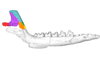
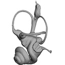
This contribution contains the 3D model described and figured in the following publication: Ramdarshan A., Orliac M.J., 2015. Endocranial morphology of Microchoerus erinaceus (Euprimates, Tarsiiformes) and early evolution of the Euprimates brain. American Journal of Physical Anthropology. doi: 10.1002/ajpa.22868
Microchoerus erinaceus UM-PRR1771 View specimen
|
M3#15Labelled 3D model of the endocranial cast and sinuse of Microchoerus erinaceus. Type: "3D_surfaces"doi: 10.18563/m3.sf15 state:published |
Download 3D surface file |
|
M3#130350µm voxel size µCT scan of the cranium of UM PRR1771 Type: "3D_CT"doi: 10.18563/m3.sf.1303 state:published |
Download CT data |
The present 3D Dataset contains the 3D models illustrated and described in the chapter “Paleoneurology of Artiodactyla, an overview of the evolution of the artiodactyl brain” (Orliac et al. 2022) published in "Paleoneurology of amniotes: new directions in the study of fossil endocasts", edited by Dozo, Paulina-Carabajal, Macrini and Walsh.
Homacodon vagans AMNH 12695 View specimen
|
M3#1063Endocranial cast Type: "3D_surfaces"doi: 10.18563/m3.sf.1063 state:published |
Download 3D surface file |
Helohyus sp. AMNH 13079 View specimen
|
M3#1064Endocranial cast Type: "3D_surfaces"doi: 10.18563/m3.sf.1064 state:published |
Download 3D surface file |
Leptauchenia sp. AMNH 45508 View specimen
|
M3#1065endocranial cast Type: "3D_surfaces"doi: 10.18563/m3.sf.1065 state:published |
Download 3D surface file |
Agriochoerus sp. AMNH 95330 View specimen
|
M3#1067endocranial cast Type: "3D_surfaces"doi: 10.18563/m3.sf.1067 state:published |
Download 3D surface file |
Mouillacitherium elegans UM ACQ 6625 View specimen
|
M3#1068endocranial cast Type: "3D_surfaces"doi: 10.18563/m3.sf.1068 state:published |
Download 3D surface file |
Caenomeryx filholi UM PDS 2570 View specimen
|
M3#1069endocranial cast Type: "3D_surfaces"doi: 10.18563/m3.sf.1069 state:published |
Download 3D surface file |
Dichobune leporina MNHN.F.QU16586 View specimen
|
M3#1070endocranial cast Type: "3D_surfaces"doi: 10.18563/m3.sf.1070 state:published |
Download 3D surface file |
Anoplotherium sp. not numbered View specimen
|
M3#1071endocranial cast Type: "3D_surfaces"doi: 10.18563/m3.sf.1071 state:published |
Download 3D surface file |
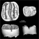
This contribution provides the raw files for the μCT-scan data and renderings of the three-dimensional digital models of two fossil teeth of a geomyin geomorph rodent (Caribeomys merzeraudi), discovered from lower Oligocene deposits of Puerto Rico, San Sebastian Formation (locality LACM Loc. 8060). These fossils were described, figured and discussed in the following publication: Marivaux et al. (2021), An unpredicted ancient colonization of the West Indies by North American rodents: dental evidence of a geomorph from the early Oligocene of Puerto Rico. Papers in Palaeontology. https://doi.org/10.1002/spp2.1388
Caribeomys merzeraudi LACM 162478 View specimen
|
M3#712Right lower dp4: isolated deciduous premolar. The specimen was scanned with a resolution of 5 µm using a μ-CT-scanning station EasyTom 150 / Rx Solutions (Montpellier RIO Imaging, ISE-M, Montpellier, France). AVIZO 7.1 (Visualization Sciences Group) software was used for visualization, segmentation, and 3D rendering. This isolated tooth was prepared within a “labelfield” module of AVIZO, using the segmentation threshold selection tool. Type: "3D_surfaces"doi: 10.18563/m3.sf.712 state:published |
Download 3D surface file |
|
M3#7145µm µCT data set . Right lower dp4: isolated deciduous premolar. The specimen was scanned with a resolution of 5 µm using a μ-CT-scanning station EasyTom 150 / Rx Solutions (Montpellier RIO Imaging, ISE-M, Montpellier, France). Type: "3D_CT"doi: 10.18563/m3.sf.714 state:published |
Download CT data |
Caribeomys merzeraudi LACM 162449 View specimen
|
M3#713Right lower molar (m1 or m2). The specimen was scanned with a resolution of 4.5 µm using a μ-CT-scanning station EasyTom 150 / Rx Solutions (Montpellier RIO Imaging, ISE-M, Montpellier, France). AVIZO 7.1 (Visualization Sciences Group) software was used for visualization, segmentation, and 3D rendering. This isolated tooth was prepared within a “labelfield” module of AVIZO, using the segmentation threshold selection tool. Type: "3D_surfaces"doi: 10.18563/m3.sf.713 state:published |
Download 3D surface file |
|
M3#715µCT data at 4.5µm Type: "3D_CT"doi: 10.18563/m3.sf.715 state:published |
Download CT data |
This contribution contains the 3D surface model of the holotype cranium of the Late Jurassic thalassochelydian turtle Solnhofia brachyrhyncha described and figured in the publication of Anquetin and Püntener (2020).
Solnhofia brachyrhyncha MJSN BAN001-2.1 View specimen
|
M3#536Textured 3D surface model of the holotype cranium of the Late Jurassic turtle Solnhofia brachyrhyncha Type: "3D_surfaces"doi: 10.18563/m3.sf.536 state:published |
Download 3D surface file |
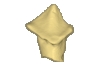
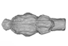
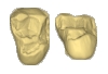
This contribution contains the 3D models of the isolated teeth of Canaanimico amazonensis, a new stem platyrrhine primate, described and figured in the following publication: Marivaux et al. (2016), Neotropics provide insights into the emergence of New World monkeys: new dental evidence from the late Oligocene of Peruvian Amazonia. Journal of Human Evolution. http://dx.doi.org/10.1016/j.jhevol.2016.05.011
Canaanimico amazonensis MUSM-2499 View specimen
|
M3#2893D model of left upper M2 Type: "3D_surfaces"doi: 10.18563/m3.sf.289 state:published |
Download 3D surface file |
Canaanimico amazonensis MUSM-2500 View specimen
|
M3#2903D model of left upper M1 (lingual part) Type: "3D_surfaces"doi: 10.18563/m3.sf.290 state:published |
Download 3D surface file |
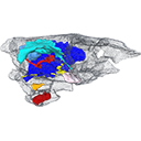
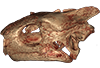
The present publication contains the µCT dataset and the 3D models analyzed in the following publication: Mautner, A.-K., A. E. Latimer, U. Fritz, and T. M. Scheyer. An updated description of the osteology of the pancake tortoise Malacochersus tornieri (Testudines: Testudinidae) with special focus on intraspecific variation. Journal of Morphology. https://doi.org/10.1002/jmor.20640
Malacochersus tornieri ZM 100.102 View specimen
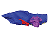
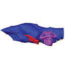
|
M3#129Virtual brain and inner ear endocast of Malacochersus tornieri (ZM 100.102; Zoological Museum of The University of Zurich). This virtual model is accompanied by the 3D dataset. Blue, endocranium; red, blood vessels; purple, semicircular canals; yellow, cranial nerves. Type: "3D_surfaces"doi: 10.18563/m3.sf.129 state:published |
Download 3D surface file |
|
M3#1303D dataset of skull of Malacochersus tornieri (ZM 100.102) Type: "3D_CT"doi: 10.18563/m3.sf.130 state:published |
Download CT data |


