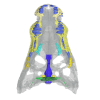
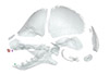
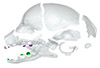
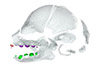
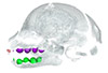
Explodable 3D Dog Skull for Veterinary Education
3D models of Ocnotherium skull
3D models of Kalakocetus, the earliest Cetacea
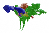
3D GM dataset of bird skeletal variation
Skeletal embryonic development in the catshark
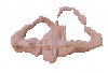
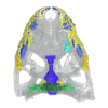
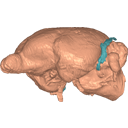
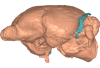
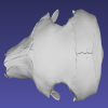
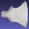
Bony connexions of the petrosal bone of extant hippos
bony labyrinth (14) , inner ear (11) , geometric morphometrics (10) , CT-scan (10) , Eocene (10) , Micro-CT (9) , Miocene (8)
Lionel Hautier (24) , Maëva Judith Orliac (23) , Laurent Marivaux (18) , Renaud Lebrun (14) , Rodolphe Tabuce (14) , Pierre-Olivier Antoine (13) , Bastien Mennecart (13)
|
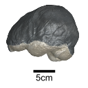
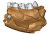
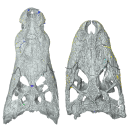
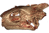
The endocranial cast of a 10 ka intentionally deformed human cranium from ChinaYin Qiyu
Published online: 27/07/2022 |
|
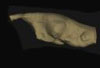
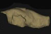
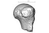
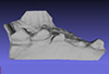
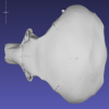
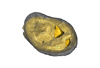
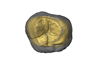
M3#972The frontal region of the endocast is flattened, probably formed by the constant pressure on the frontal bone during growth. There is a well-developed frontal crest on the endocranial surface. The endocast widens posteriorly from the frontal lobe. The widest point of the endocast is at the lateral border of the parietal lobe. The lower parietal areas display a marked lateral expansion. The overall shape of the endocast is asymmetrical, with the left side of the parietal lobe being more laterally expanded than the right side. Like the frontal lobe, the occipital lobe is also anteroposteriorly flattened. Type: "3D_surfaces"doi: 10.18563/m3.sf.972 state:published |
Download 3D surface file |
|
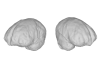
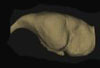
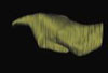
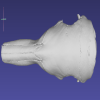
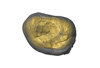
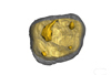
M3#976The original endocranial cast model (with texture) of IVPP-PA1616. It shows the original structures of the specimen, and was not altered in any way. Type: "3D_surfaces"doi: 10.18563/m3.sf.976 state:published |
Download 3D surface file |
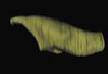
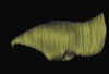
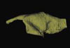
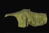
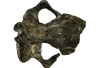
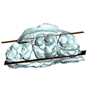
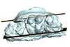
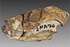
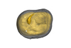
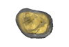
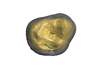
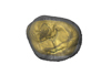
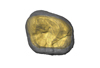
The present 3D dataset contains the 3D models of the holotype of Proterochampsa nodosa that were built and analysed in “Redescription, taxonomic revaluation, and phylogenetic affinities of Proterochampsa nodosa (Archosauriformes: Proterochampsidae), early Late Triassic of Candelaria Sequence (Santa Maria Supersequence)”.
Proterochampsa nodosa MCP 1694-PV View specimen
|
M3#9743D models of Proterochampsa nodosa. Model 1: skull. Model 2: mandible. Model 3: left mandibular ramus. Type: "3D_surfaces"doi: 10.18563/m3.sf.974 state:published |
Download 3D surface file |
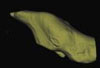
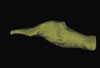
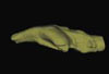
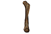
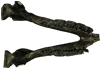
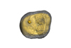
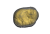
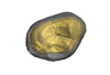
This note presents the 3D model of the hemi-mandible UM-PAT 159 of the MP7 Diacodexis species D. cf. gigasei and 3D models corresponding to the restoration of the ascending ramus, broken on the original specimen, and to a restoration of a complete mandible based on the preserved left hemi-mandible.
Diacodexis cf. gigasei UMPAT159 View specimen
|
M3#3153D models of UM PAT 159 after the restoration of the ascending ramus Type: "3D_surfaces"doi: 10.18563/m3.sf.315 state:published |
Download 3D surface file |
|
M3#316restoration of a complete mandible based on the preserved left hemi-mandible UM PAT 159 Type: "3D_surfaces"doi: 10.18563/m3.sf.316 state:published |
Download 3D surface file |
|
M3#3173D model of the hemi-mandible UM PAT 159 Type: "3D_surfaces"doi: 10.18563/m3.sf.317 state:published |
Download 3D surface file |
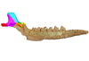
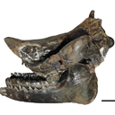
The present 3D Dataset contains the 3D models analyzed in Pochat-Cottilloux Y., Rinder N., Perrichon G., Adrien J., Amiot R., Hua S. & Martin J. E. (2023). The neuroanatomy and pneumaticity of Hamadasuchus from the Cretaceous of Morocco and its significance for the paleoecology of Peirosauridae and other altirostral crocodylomorphs. Journal of Anatomy, https://doi.org/10.1111/joa.13887
Hamadasuchus sp. UCBL-FSL 532408 View specimen
|
M3#10943D volume reconstruction of the braincase osteology Type: "3D_surfaces"doi: 10.18563/m3.sf.1094 state:published |
Download 3D surface file |
|
M3#10963D volume reconstruction of the endocast Type: "3D_surfaces"doi: 10.18563/m3.sf.1096 state:published |
Download 3D surface file |
|
M3#10973D volume reconstruction of the labyrinths Type: "3D_surfaces"doi: 10.18563/m3.sf.1097 state:published |
Download 3D surface file |
|
M3#10983D volume reconstruction of the pneumatic cavities Type: "3D_surfaces"doi: 10.18563/m3.sf.1098 state:published |
Download 3D surface file |
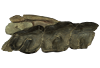
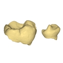
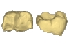
The present 3D Dataset contains the 3D models analyzed in Mennecart B., Wazir W.A., Sehgal R.K., Patnaik R., Singh N.P., Kumar N, and Nanda A.C. 2021. New remains of Nalamaeryx (Tragulidae, Mammalia) from the Ladakh Himalaya and their phylogenetical and palaeoenvironmental implications. Historical Biology. https://doi.org/10.1080/08912963.2021.2014479
Nalameryx savagei WIMF/A4801 View specimen
|
M3#766Nalameryx savagei, Partial lower right jaw preserving m2 and m3. Type: "3D_surfaces"doi: 10.18563/m3.sf.766 state:published |
Download 3D surface file |
Nalameryx savagei WIMF/A4802 View specimen
|
M3#767Nalameryx savagei, partial lower right jaw preserving m2 and m3 Type: "3D_surfaces"doi: 10.18563/m3.sf.767 state:published |
Download 3D surface file |
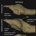
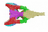
The present 3D Dataset contains 26 3D models analyzed in the study: On the “cartilaginous rider” in the endocasts of turtle brain cavities, published by the authors in the journal Vertebrate Zoology.
Annemys sp. IVPP-V-18106 View specimen

|
M3#7723D surface(s) file to specimen IVPP-V-18106 Type: "3D_surfaces"doi: 10.18563/m3.sf.772 state:published |
Download 3D surface file |
Apalone spinifera FMNH 22178 View specimen

|
M3#7733D surface(s) file to specimen FMNH 22178 Type: "3D_surfaces"doi: 10.18563/m3.sf.773 state:published |
Download 3D surface file |
Caretta caretta NHMUK1940.3.15.1 View specimen
|
M3#7863D surface(s) file to specimen NHMUK1940.3.15.1 Type: "3D_surfaces"doi: 10.18563/m3.sf.786 state:published |
Download 3D surface file |
Chelodina reimanni ZMB 49659 View specimen

|
M3#7743D surface(s) file to specimen ZMB 49659 Type: "3D_surfaces"doi: 10.18563/m3.sf.774 state:published |
Download 3D surface file |
Chelonia mydas ZMB-37416MS View specimen
|
M3#7753D surface(s) file to specimen ZMB-37416MS Type: "3D_surfaces"doi: 10.18563/m3.sf.775 state:published |
Download 3D surface file |
Cuora amboinensis NHMUK69.42.145_4 View specimen
|
M3#7763D surface(s) file to specimen NHMUK69.42.145_4 Type: "3D_surfaces"doi: 10.18563/m3.sf.776 state:published |
Download 3D surface file |
Emydura subglobosa IW92 View specimen
|
M3#7773D surface(s) file to specimen IW92 Type: "3D_surfaces"doi: 10.18563/m3.sf.777 state:published |
Download 3D surface file |
Eubaena cephalica DMNH 96004 View specimen
|
M3#7783D surface(s) file to specimen DMNH 96004 Type: "3D_surfaces"doi: 10.18563/m3.sf.778 state:published |
Download 3D surface file |
Gopherus berlandieri AMNH-73816 View specimen
|
M3#7793D surface(s) file to specimen AMNH-73816 Type: "3D_surfaces"doi: 10.18563/m3.sf.779 state:published |
Download 3D surface file |
Kinixys belliana AMNH-10028 View specimen
|
M3#7803D surface(s) file to specimen AMNH-10028 Type: "3D_surfaces"doi: 10.18563/m3.sf.780 state:published |
Download 3D surface file |
Macrochelys temminckii GPIT-PV-79430 View specimen

|
M3#7813D surface(s) file to specimen GPIT-PV-79430 Type: "3D_surfaces"doi: 10.18563/m3.sf.781 state:published |
Download 3D surface file |
Malacochersus tornieri SMF-58702 View specimen
|
M3#7873D surface(s) file to specimen SMF-58702 Type: "3D_surfaces"doi: 10.18563/m3.sf.787 state:published |
Download 3D surface file |
Naomichelys speciosa FMNH-PR-273 View specimen
|
M3#7823D surface(s) file to specimen FMNH-PR-273 Type: "3D_surfaces"doi: 10.18563/m3.sf.782 state:published |
Download 3D surface file |
Pelodiscus sinensis IW576-2 View specimen
|
M3#7833D surface(s) file to specimen IW576-2 Type: "3D_surfaces"doi: 10.18563/m3.sf.783 state:published |
Download 3D surface file |
Platysternon megacephalum SMF-69684 View specimen
|
M3#7843D surface(s) file to specimen SMF-69684 Type: "3D_surfaces"doi: 10.18563/m3.sf.784 state:published |
Download 3D surface file |
Podocnemis unifilis SMF-55470 View specimen
|
M3#7853D surface(s) file to specimen SMF-55470 Type: "3D_surfaces"doi: 10.18563/m3.sf.785 state:published |
Download 3D surface file |
Proganochelys quenstedtii MB 1910.45.2 View specimen
|
M3#7883D surface(s) file to specimen MB 1910.45.2 Type: "3D_surfaces"doi: 10.18563/m3.sf.788 state:published |
Download 3D surface file |
Proganochelys quenstedtii SMNS 16980 View specimen
|
M3#7893D surface(s) file to specimen SMNS 16980 Type: "3D_surfaces"doi: 10.18563/m3.sf.789 state:published |
Download 3D surface file |
Rhinochelys pulchriceps CAMSM_B55775 View specimen
|
M3#7903D surface(s) file to specimen CAMSM_B55775 Type: "3D_surfaces"doi: 10.18563/m3.sf.790 state:published |
Download 3D surface file |
Rhinoclemmys funereal YPM12174 View specimen
|
M3#7913D surface(s) file to specimen YPM12174 Type: "3D_surfaces"doi: 10.18563/m3.sf.791 state:published |
Download 3D surface file |
Sandownia harrisi MIWG3480 View specimen
|
M3#7923D surface(s) file to specimen MIWG3480 Type: "3D_surfaces"doi: 10.18563/m3.sf.792 state:published |
Download 3D surface file |
Testudo graeca YPM14342 View specimen
|
M3#7933D surface(s) file to specimen YPM14342 Type: "3D_surfaces"doi: 10.18563/m3.sf.793 state:published |
Download 3D surface file |
Testudo hermanni AMNH134518 View specimen
|
M3#7943D surface(s) file to specimen AMNH134518 Type: "3D_surfaces"doi: 10.18563/m3.sf.794 state:published |
Download 3D surface file |
Trachemys scripta NN View specimen
|
M3#7953D surface(s) file to specimen Trachemys scripta Type: "3D_surfaces"doi: 10.18563/m3.sf.795 state:published |
Download 3D surface file |
Xinjiangchelys radiplicatoides IVPP V9539 View specimen
|
M3#7963D surface(s) file to specimen IVPP V9539 Type: "3D_surfaces"doi: 10.18563/m3.sf.796 state:published |
Download 3D surface file |
Chelydra serpentina UFR VP1 View specimen

|
M3#801Brain endocast Type: "3D_surfaces"doi: 10.18563/m3.sf.801 state:published |
Download 3D surface file |
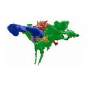
This contribution contains the 3D models of the paratympanic sinus system, the endocast and the neurovascular bony canal of the maxilla, premaxilla and the jugal of Leidyosuchus canadensis and Stangerochampsa mccabei described and figured in the following publication: G. Donzé, G. Perrichon, P. Vincent, JE. Martin, 2026. Comparative endocranial traits in the crocodylians Leidyosuchus canadensis and Stangerochampsa mccabei from the upper Cretaceous of Alberta, Canada. Journal of Anatomy.
Leidyosuchus canadensis TMP1986.221.1 View specimen
|
M3#1830Skull, endocast, paratympanic sinuses, jugal and maxillary neurovascular canals, alveoli Type: "3D_surfaces"doi: 10.18563/m3.sf.1830 state:in_press |
Download 3D surface file |
Stangerochampsa mccabei TMP1986.61.1 View specimen
|
M3#1831Skull, endocast, paratympanic sinuses, jugal and maxillary neurovascular canals, alveoli Type: "3D_surfaces"doi: 10.18563/m3.sf.1831 state:in_press |
Download 3D surface file |
Alligator mississipiensis OUVC 9761 View specimen

|
M3#1833Skull, endocast, paratympanic sinuses, jugal and maxillary neurovascular canals, alveolies Type: "3D_surfaces"doi: 10.18563/m3.sf.1833 state:in_press |
Download 3D surface file |
Alligator sinensis NHMW-Zoo-HS-37966 View specimen
|
M3#1835Skull, endocast, paratympanic sinus, jugal and maxillary neurovascular canals, alveoli Type: "3D_surfaces"doi: 10.18563/m3.sf.1835 state:in_press |
Download 3D surface file |
Crocodylus niloticus MHNL 50001399 View specimen
|
M3#1834Skull, endocast, paratympanic sinuses, jugal and maxillary neurovascular canals, alveolies Type: "3D_surfaces"doi: 10.18563/m3.sf.1834 state:in_press |
Download 3D surface file |
Diplocynodon ratelii LA86 View specimen
|
M3#1832Skull, endocast, paratympanic sinus, jugal and maxillary neurovascular canals, alveolies Type: "3D_surfaces"doi: 10.18563/m3.sf.1832 state:in_press |
Download 3D surface file |
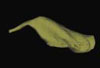
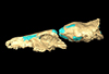
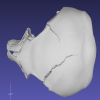
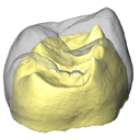
This contribution contains the 3D model described and figured in the following publication: Ramdarshan A., Orliac M.J., 2015. Endocranial morphology of Microchoerus erinaceus (Euprimates, Tarsiiformes) and early evolution of the Euprimates brain. American Journal of Physical Anthropology. doi: 10.1002/ajpa.22868
Microchoerus erinaceus UM-PRR1771 View specimen
|
M3#15Labelled 3D model of the endocranial cast and sinuse of Microchoerus erinaceus. Type: "3D_surfaces"doi: 10.18563/m3.sf15 state:published |
Download 3D surface file |
|
M3#130350µm voxel size µCT scan of the cranium of UM PRR1771 Type: "3D_CT"doi: 10.18563/m3.sf.1303 state:published |
Download CT data |
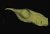
This contribution contains the 3D model described and figured in the following publication: Dubied, M., Mennecart, B. and Solé, F. 2019. The cranium of Proviverra typica (Mammalia, Hyaenodonta) and its impact on hyaenodont phylogeny and endocranial evolution. Palaeontology. https://doi.org/10.1111/pala.12437
Proviverra typica NMB Em18 View specimen
|
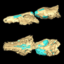
M3#355The file contain the cranium (yellow) and the endocast (blue) of the facial part and the brain case part of the type specimen of Proviverra typica (NMB Em18). Type: "3D_surfaces"doi: 10.18563/m3.sf.355 state:published |
Download 3D surface file |
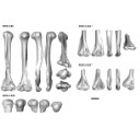
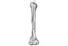
The present 3D Dataset contains the 3D models analyzed in the publication “Systematic and locomotor diversification of the Adapis group (Primates, Adapiformes) in the late Eocene of the Quercy (Southwest France), revealed by humeral remains”. In this paper, twenty humeral specimens from the old and new Quercy collections attributed to the fossil primates Adapis and Palaeolemur are described and analysed together. In this dataset only the scans of the fossils belonging to the collections of Université de Montpellier are provided.
In our paper (Marigó et al., 2019) we provide a qualitative and quantitative analysis of the different humeri, revealing that high variability is present within the “Adapis group” sample. Six different morphotypes are identified, confirming that what has often been called “Adapis parisiensis” is a mix of different species that present different locomotor adaptations.
Adapis sp. UM ROS 2-95 View specimen
|
M3#356Complete right humerus ROS 2-95 attributed to the Adapis group Type: "3D_surfaces"doi: 10.18563/m3.sf.356 state:published |
Download 3D surface file |
Adapis sp. UM ROS 2-536 View specimen
|
M3#357Proximal end of the right humerus ROS 2-536 attributed to the Adapis group Type: "3D_surfaces"doi: 10.18563/m3.sf.357 state:published |
Download 3D surface file |
Adapis sp. UM ROS 2-534 View specimen
|
M3#358Distal end of the left humerus ROS 2-534 attributed to the Adapis group Type: "3D_surfaces"doi: 10.18563/m3.sf.358 state:published |
Download 3D surface file |
Adapis sp. UM ROS 2-535 View specimen
|
M3#359Distal end of the left humerus ROS 2-535 attributed to the Adapis group Type: "3D_surfaces"doi: 10.18563/m3.sf.359 state:published |
Download 3D surface file |
Adapis sp. UM ROS 2-80 View specimen

|
M3#360Proximal end of the right humerus ROS 2-80 attributed to the Adapis group Type: "3D_surfaces"doi: 10.18563/m3.sf.360 state:published |
Download 3D surface file |
Adapis sp. UM ROS 2-79 View specimen
|
M3#361Distal end of the right humerus ROS 2-79 attributed to the Adapis group Type: "3D_surfaces"doi: 10.18563/m3.sf.361 state:published |
Download 3D surface file |
Adapis sp. UM ECA 1364 View specimen

|
M3#362Distal end of the left humerus ECA 1364 attributed to the Adapis group Type: "3D_surfaces"doi: 10.18563/m3.sf.362 state:published |
Download 3D surface file |
Adapis sp. UM ACQ-262 View specimen
|
M3#3733D model of ACQ 262. Humerus Type: "3D_surfaces"doi: 10.18563/m3.sf373 state:published |
Download 3D surface file |
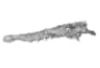
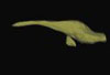
This contribution contains the 3D model described and figured in the following publication: Albino, A., Carrillo-Briceño, J. D. & Neenan, J. M. 2016. An enigmatic aquatic snake from the Cenomanian of northern South America. PeerJ 4:e2027 http://dx.doi.org/10.7717/peerj.2027
Lunaophis aquaticus MCNC-1827-F View specimen
|
M3#116Articulated precloacal vertebrae of Lunaophis aquaticus Type: "3D_surfaces"doi: 10.18563/m3.sf.116 state:published |
Download 3D surface file |
This contribution contains the 3D models described and figured in the following publication: Tissier et al. (in prep.).
Sellamynodon zimborensis UBB MPS 15795 View specimen
|
M3#297Incomplete skull with left M3. Type: "3D_surfaces"doi: 10.18563/m3.sf.297 state:published |
Download 3D surface file |
Sellamynodon zimborensis UBB MPS 15795 View specimen
|
M3#298Mandible with complete molar and premolar rows, lacking symphysis. Type: "3D_surfaces"doi: 10.18563/m3.sf.298 state:published |
Download 3D surface file |
Amynodontopsis aff. bodei UBB MPS V545 View specimen
|
M3#299Maxillary fragment with M1-3. Type: "3D_surfaces"doi: 10.18563/m3.sf.299 state:published |
Download 3D surface file |
Amynodontopsis aff. bodei UBB MPS V546 View specimen
|
M3#300Unworn m1/2 on mandible fragment. Type: "3D_surfaces"doi: 10.18563/m3.sf.300 state:published |
Download 3D surface file |
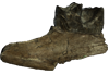
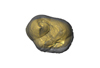
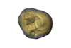
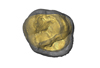
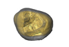
This contribution contains the 3D models described and figured in the following publication: Orliac M.J., Karadenizli L., Antoine P.-O., Sen S. 2015. Small suids (Mammalia, Artiodactyla) from the late Early Miocene of Turkey and a short overview of Early Miocene small suoids in the Old World. Paleontologia electronica 18(2): 18.2.30A: 1-48. https://doi.org/10.26879/547
?Nguruwe galaticum SMT-1 View specimen
|
M3#16fragment of palate with left broken M1-M3 Type: "3D_surfaces"doi: 10.18563/m3.sf16 state:published |
Download 3D surface file |
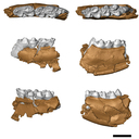
The present 3D Dataset contains the 3D models analyzed in Hendrickx, C., Gaetano, L. C., Choiniere, J., Mocke, H. and Abdala, F. in press. A new traversodontid cynodont with a peculiar postcanine dentition from the Middle/Late Triassic of Namibia and dental evolution in basal gomphodonts. Journal of Systematic Palaeontology.
Etjoia dentitransitus GSN F1591 View specimen
|
M3#557Surface model derived from µCT data of the holotype of Etjoia dentitransitus Type: "3D_surfaces"doi: 10.18563/m3.sf.557 state:published |
Download 3D surface file |
|
M3#558Photogrammetric 3D surface model of the postcanines of the Holotype of Etjoia dentitransitus Type: "3D_surfaces"doi: 10.18563/m3.sf.558 state:published |
Download 3D surface file |
|
M3#559Photogrammetric 3D surface model of the Holotype of Etjoia dentitransitus Type: "3D_surfaces"doi: 10.18563/m3.sf.559 state:published |
Download 3D surface file |
This contribution contains the 3D models described and figured in: Phylogenetic signal in anteater snout morphology: implications for interpreting rare vermilinguan fossils. Palaeobiodiversity and Palaeoenvironments.
Indet indet VPPLT 977 View specimen

|
M3#17933D surface models of the cranium, nasal bone and cranial canals Type: "3D_surfaces"doi: 10.18563/m3.sf.1793 state:in_press |
Download 3D surface file |
Cyclopes didactylus M 1525 View specimen
|
M3#17943D models of the cranium and internal cranial canals Type: "3D_surfaces"doi: 10.18563/m3.sf.1794 state:in_press |
Download 3D surface file |
Cyclopes didactylus M 1571 View specimen
|
M3#17953D surface models of the cranium, nasal bone and cranial canals Type: "3D_surfaces"doi: 10.18563/m3.sf.1795 state:in_press |
Download 3D surface file |
Myrmecophaga tridactyla M 3023 View specimen
|
M3#17963D surface models of the cranium, nasal bone and cranial canals Type: "3D_surfaces"doi: 10.18563/m3.sf.1796 state:in_press |
Download 3D surface file |
Tamandua tetradactyla NHMUK 3.7.7.135 View specimen
|
M3#17973D models of the cranium and internal cranial canals Type: "3D_surfaces"doi: 10.18563/m3.sf.1797 state:in_press |
Download 3D surface file |
Tamandua tetradactyla NHMUK 4.7.4.90 View specimen
|
M3#17983D surface models of the cranium, nasal bone and cranial canals Type: "3D_surfaces"doi: 10.18563/m3.sf.1798 state:in_press |
Download 3D surface file |
Tamandua tetradactyla UM 788N View specimen
|
M3#17993D models of the cranium and internal cranial canals Type: "3D_surfaces"doi: 10.18563/m3.sf.1799 state:in_press |
Download 3D surface file |
Cyclopes didactylus MVZ 121210 View specimen
|
M3#18003D models of the cranium and internal cranial canals Type: "3D_surfaces"doi: 10.18563/m3.sf.1800 state:in_press |
Download 3D surface file |
Myrmecophaga tridactyla MVZ 112943 View specimen
|
M3#18013D models of the cranium and internal cranial canals Type: "3D_surfaces"doi: 10.18563/m3.sf.1801 state:in_press |
Download 3D surface file |
Myrmecophaga tridactyla MVZ 185238 View specimen
|
M3#18023D models of the cranium and internal cranial canals Type: "3D_surfaces"doi: 10.18563/m3.sf.1802 state:in_press |
Download 3D surface file |
The present 3D Dataset contains the 3D models of external and internal aspects of human upper permanent second molars from the Neolithic necropolis analyzed in the following publication: Le Luyer M., Coquerelle M., Rottier S., Bayle P. (2016): Internal tooth structure and burial practices: insights into the Neolithic necropolis of Gurgy (France, 5100-4000 cal. BC). Plos One 11(7): e0159688. doi: 10.1371/journal.pone.0159688.
Homo sapiens GLN04-201-ULM2 View specimen
|
M3#74Outer enamel surface (OES) and enamel-dentine junction (EDJ) of Neolithic upper permanent left second molar Type: "3D_surfaces"doi: 10.18563/m3.sf.74 state:published |
Download 3D surface file |
Homo sapiens GLN04-206-ULM2 View specimen
|
M3#75Outer enamel surface (OES) and enamel-dentine junction (EDJ) of Neolithic upper permanent left second molar Type: "3D_surfaces"doi: 10.18563/m3.sf.75 state:published |
Download 3D surface file |
Homo sapiens GLN05-213-URM2 View specimen
|
M3#76Outer enamel surface (OES) and enamel-dentine junction (EDJ) of Neolithic upper permanent right second molar Type: "3D_surfaces"doi: 10.18563/m3.sf.76 state:published |
Download 3D surface file |
Homo sapiens GLN05-215A-URM2 View specimen
|
M3#77Outer enamel surface (OES) and enamel-dentine junction (EDJ) of Neolithic upper permanent right second molar Type: "3D_surfaces"doi: 10.18563/m3.sf.77 state:published |
Download 3D surface file |
Homo sapiens GLN06-215B-URM2 View specimen
|
M3#78Outer enamel surface (OES) and enamel-dentine junction (EDJ) of Neolithic upper permanent right second molar Type: "3D_surfaces"doi: 10.18563/m3.sf.78 state:published |
Download 3D surface file |
Homo sapiens GLN06-223-URM2 View specimen
|
M3#79Outer enamel surface (OES) and enamel-dentine junction (EDJ) of Neolithic upper permanent right second molar Type: "3D_surfaces"doi: 10.18563/m3.sf.79 state:published |
Download 3D surface file |
Homo sapiens GLN04-229-URM2 View specimen
|
M3#80Outer enamel surface (OES) and enamel-dentine junction (EDJ) of Neolithic upper permanent right second molar Type: "3D_surfaces"doi: 10.18563/m3.sf.80 state:published |
Download 3D surface file |
Homo sapiens GLN05-243B-ULM2 View specimen
|
M3#81Outer enamel surface (OES) and enamel-dentine junction (EDJ) with reconstructed dentine horn tip of Neolithic upper permanent left second molar Type: "3D_surfaces"doi: 10.18563/m3.sf.81 state:published |
Download 3D surface file |
Homo sapiens GLN04-248-ULM2 View specimen
|
M3#82Outer enamel surface (OES) and enamel-dentine junction (EDJ) with reconstructed dentine horn tip of Neolithic upper permanent left second molar Type: "3D_surfaces"doi: 10.18563/m3.sf.82 state:published |
Download 3D surface file |
Homo sapiens GLN04-252-ULM2 View specimen
|
M3#83Outer enamel surface (OES) and enamel-dentine junction (EDJ) of Neolithic upper permanent left second molar Type: "3D_surfaces"doi: 10.18563/m3.sf.83 state:published |
Download 3D surface file |
Homo sapiens GLN04-253-ULM2 View specimen
|
M3#84Outer enamel surface (OES) and enamel-dentine junction (EDJ) of Neolithic upper permanent left second molar Type: "3D_surfaces"doi: 10.18563/m3.sf.84 state:published |
Download 3D surface file |
Homo sapiens GLN05-257-URM2 View specimen
|
M3#85Outer enamel surface (OES) and enamel-dentine junction (EDJ) with reconstructed dentine horn tip of Neolithic upper permanent right second molar Type: "3D_surfaces"doi: 10.18563/m3.sf.85 state:published |
Download 3D surface file |
Homo sapiens GLN04-264-ULM2 View specimen
|
M3#86Outer enamel surface (OES) and enamel-dentine junction (EDJ) of Neolithic upper permanent left second molar Type: "3D_surfaces"doi: 10.18563/m3.sf.86 state:published |
Download 3D surface file |
Homo sapiens GLN04-277-URM2 View specimen
|
M3#87Outer enamel surface (OES) and enamel-dentine junction (EDJ) of Neolithic upper permanent right second molar Type: "3D_surfaces"doi: 10.18563/m3.sf.87 state:published |
Download 3D surface file |
Homo sapiens GLN04-289B-URM2 View specimen
|
M3#88Outer enamel surface (OES) and enamel-dentine junction (EDJ) of Neolithic upper permanent right second molar Type: "3D_surfaces"doi: 10.18563/m3.sf.88 state:published |
Download 3D surface file |
Homo sapiens GLN06-291-URM2 View specimen
|
M3#89Outer enamel surface (OES) and enamel-dentine junction (EDJ) with reconstructed dentine horn tip of Neolithic upper permanent right second molar Type: "3D_surfaces"doi: 10.18563/m3.sf.89 state:published |
Download 3D surface file |
Homo sapiens GLN05-292-URM2 View specimen
|
M3#90Outer enamel surface (OES) and enamel-dentine junction (EDJ) of Neolithic upper permanent right second molar Type: "3D_surfaces"doi: 10.18563/m3.sf.90 state:published |
Download 3D surface file |
Homo sapiens GLN05-294-ULM2 View specimen
|
M3#91Outer enamel surface (OES) and enamel-dentine junction (EDJ) with reconstructed dentine horn tip of Neolithic upper permanent left second molar Type: "3D_surfaces"doi: 10.18563/m3.sf.91 state:published |
Download 3D surface file |
Homo sapiens GLN05-308-URM2 View specimen
|
M3#93Outer enamel surface (OES) and enamel-dentine junction (EDJ) of Neolithic upper permanent right second molar Type: "3D_surfaces"doi: 10.18563/m3.sf.93 state:published |
Download 3D surface file |
Homo sapiens GLN05-301-ULM2 View specimen
|
M3#92Outer enamel surface (OES) and enamel-dentine junction (EDJ) of Neolithic upper permanent left second molar Type: "3D_surfaces"doi: 10.18563/m3.sf.92 state:published |
Download 3D surface file |
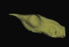
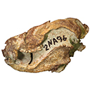
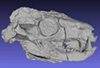
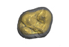
This contribution contains the 3D model of the holotype of Chambius kasserinensis, the basalmost ‘elephant-shrew’ figured in the following publication: New remains of Chambius kasserinensis from the Eocene of Tunisia and evaluation of proposed affinities for Macroscelidea (Mammalia, Afrotheria). https://doi.org/10.1080/08912963.2017.1297433
Chambius kasserinensis CBI-1-06 View specimen
|
M3#1463D model of the holotype maxilla of Chambius kasserinensis. The 3D surface was extracted manually from the limestone matrix within AVIZO 9.2 Type: "3D_surfaces"doi: 10.18563/m3.sf.146 state:published |
Download 3D surface file |
This contribution contains the 3D models described and figured in the following publication: Hautier L., Gomes Rodrigues H., Billet G., Asher R.J., 2016. The hidden teeth of sloths: evolutionary vestiges and the development of a simplified dentition. Scientific Reports. doi: 10.1038/srep27763
Bradypus variegatus ZMB 33812 View specimen
|
M3#110Three-dimensional reconstruction of the teeth, mandibles, maxillary and premaxillary bones Type: "3D_surfaces"doi: 10.18563/m3.sf.110 state:published |
Download 3D surface file |
Bradypus variegatus ZMB 41122 View specimen
|
M3#109Three-dimensional reconstruction of the teeth, mandibles, maxillary and premaxillary bones Type: "3D_surfaces"doi: 10.18563/m3.sf.109 state:published |
Download 3D surface file |
Bradypus variegatus MNHN-ZM-MO-1995-326A View specimen
|
M3#111Three-dimensional reconstruction of the teeth, mandibles, maxillary and premaxillary bones Type: "3D_surfaces"doi: 10.18563/m3.sf.111 state:published |
Download 3D surface file |
Bradypus variegatus MNHN-ZM-MO-1995-326B View specimen
|
M3#112Three-dimensional reconstruction of the teeth, mandibles, maxillary and premaxillary bones Type: "3D_surfaces"doi: 10.18563/m3.sf.112 state:published |
Download 3D surface file |
Bradypus sp. MNHN-ZM-MO-1902-325 View specimen
|
M3#113Three-dimensional reconstruction of the teeth, mandibles, maxillary, and premaxillary bones Type: "3D_surfaces"doi: 10.18563/m3.sf.113 state:published |
Download 3D surface file |
Bradypus sp. MNHN-ZM-MO-1995-327 View specimen
|
M3#114Three-dimensional reconstruction of the teeth, mandibles, maxillary and premaxillary bones Type: "3D_surfaces"doi: 10.18563/m3.sf.114 state:published |
Download 3D surface file |
Choloepus didactylus MNHN-ZM-MO-1882-625 View specimen
|
M3#115Three-dimensional reconstruction of the teeth, mandibles, maxillary and premaxillary bones Type: "3D_surfaces"doi: 10.18563/m3.sf.115 state:published |
Download 3D surface file |
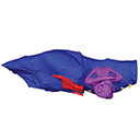
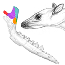
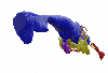
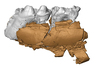
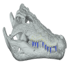
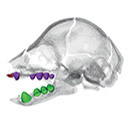
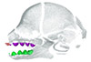
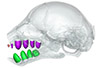
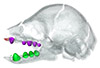
The present publication contains the µCT dataset and the 3D models analyzed in the following publication: Mautner, A.-K., A. E. Latimer, U. Fritz, and T. M. Scheyer. An updated description of the osteology of the pancake tortoise Malacochersus tornieri (Testudines: Testudinidae) with special focus on intraspecific variation. Journal of Morphology. https://doi.org/10.1002/jmor.20640
Malacochersus tornieri ZM 100.102 View specimen

|
M3#129Virtual brain and inner ear endocast of Malacochersus tornieri (ZM 100.102; Zoological Museum of The University of Zurich). This virtual model is accompanied by the 3D dataset. Blue, endocranium; red, blood vessels; purple, semicircular canals; yellow, cranial nerves. Type: "3D_surfaces"doi: 10.18563/m3.sf.129 state:published |
Download 3D surface file |
|
M3#1303D dataset of skull of Malacochersus tornieri (ZM 100.102) Type: "3D_CT"doi: 10.18563/m3.sf.130 state:published |
Download CT data |
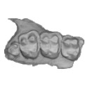
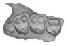
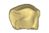
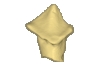
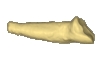
This contribution contains the 3D models of the isolated teeth attributed to stem representatives of the Cebuella and Cebus lineages (Cebuella sp. and Cebus sp.), described and figured in the following publication: Marivaux et al. (2016), Dental remains of cebid platyrrhines from the earliest late Miocene of Western Amazonia, Peru: macroevolutionary implications on the extant capuchin and marmoset lineages. American Journal of Physical Anthropology. http://dx.doi.org/10.1002/ajpa.23052
Cebus sp. MUSM-3243 View specimen
|
M3#2823D model of left lower m1 (lingual part) Type: "3D_surfaces"doi: 10.18563/m3.sf.282 state:published |
Download 3D surface file |
Cebuella sp. MUSM-3239 View specimen
|
M3#2833D model of left lower p4 Type: "3D_surfaces"doi: 10.18563/m3.sf.283 state:published |
Download 3D surface file |
Cebuella sp. MUSM-3240 View specimen
|
M3#2943D model of right upper P3 or P4 (buccal part) Type: "3D_surfaces"doi: 10.18563/m3.sf.294 state:published |
Download 3D surface file |
Cebuella sp. MUSM-3241 View specimen
|
M3#2953D model of right upper P2 Type: "3D_surfaces"doi: 10.18563/m3.sf.295 state:published |
Download 3D surface file |
Cebuella sp. MUSM-3242 View specimen
|
M3#2963D model of upper I2 Type: "3D_surfaces"doi: 10.18563/m3.sf.296 state:published |
Download 3D surface file |


