Explodable 3D Dog Skull for Veterinary Education
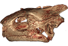
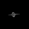
3D models of Ocnotherium skull
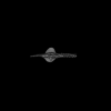
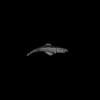
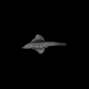
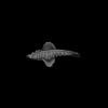
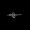
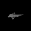
3D models of Kalakocetus, the earliest Cetacea
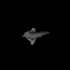
3D GM dataset of bird skeletal variation
Skeletal embryonic development in the catshark
Bony connexions of the petrosal bone of extant hippos
bony labyrinth (14) , inner ear (11) , geometric morphometrics (10) , CT-scan (10) , Eocene (10) , Micro-CT (9) , Miocene (8)
Lionel Hautier (24) , Maëva Judith Orliac (23) , Laurent Marivaux (18) , Renaud Lebrun (14) , Rodolphe Tabuce (14) , Pierre-Olivier Antoine (13) , Bastien Mennecart (13)
|
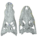
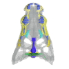
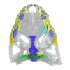
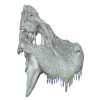
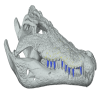
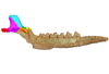
3D models related to the publication: Comparative endocranial traits in the crocodylians Leidyosuchus canadensis and Stangerochampsa mccabei from the upper Cretaceous of Alberta, Canada.Garance Donzé
Published online: 14/01/2026 |
|
M3#1830Skull, endocast, paratympanic sinuses, jugal and maxillary neurovascular canals, alveoli Type: "3D_surfaces"doi: 10.18563/m3.sf.1830 state:in_press |
Download 3D surface file |
Stangerochampsa mccabei TMP1986.61.1 View specimen
|
M3#1831Skull, endocast, paratympanic sinuses, jugal and maxillary neurovascular canals, alveoli Type: "3D_surfaces"doi: 10.18563/m3.sf.1831 state:in_press |
Download 3D surface file |
Alligator mississipiensis OUVC 9761 View specimen

|
M3#1833Skull, endocast, paratympanic sinuses, jugal and maxillary neurovascular canals, alveolies Type: "3D_surfaces"doi: 10.18563/m3.sf.1833 state:in_press |
Download 3D surface file |
Alligator sinensis NHMW-Zoo-HS-37966 View specimen
|
M3#1835Skull, endocast, paratympanic sinus, jugal and maxillary neurovascular canals, alveoli Type: "3D_surfaces"doi: 10.18563/m3.sf.1835 state:in_press |
Download 3D surface file |
Crocodylus niloticus MHNL 50001399 View specimen
|
M3#1834Skull, endocast, paratympanic sinuses, jugal and maxillary neurovascular canals, alveolies Type: "3D_surfaces"doi: 10.18563/m3.sf.1834 state:in_press |
Download 3D surface file |
Diplocynodon ratelii LA86 View specimen
|
M3#1832Skull, endocast, paratympanic sinus, jugal and maxillary neurovascular canals, alveolies Type: "3D_surfaces"doi: 10.18563/m3.sf.1832 state:in_press |
Download 3D surface file |
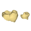
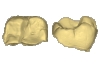
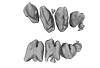
This contribution contains the 3D models of the isolated teeth attributed to stem representatives of the Cebuella and Cebus lineages (Cebuella sp. and Cebus sp.), described and figured in the following publication: Marivaux et al. (2016), Dental remains of cebid platyrrhines from the earliest late Miocene of Western Amazonia, Peru: macroevolutionary implications on the extant capuchin and marmoset lineages. American Journal of Physical Anthropology. http://dx.doi.org/10.1002/ajpa.23052
Cebus sp. MUSM-3243 View specimen
|
M3#2823D model of left lower m1 (lingual part) Type: "3D_surfaces"doi: 10.18563/m3.sf.282 state:published |
Download 3D surface file |
Cebuella sp. MUSM-3239 View specimen
|
M3#2833D model of left lower p4 Type: "3D_surfaces"doi: 10.18563/m3.sf.283 state:published |
Download 3D surface file |
Cebuella sp. MUSM-3240 View specimen
|
M3#2943D model of right upper P3 or P4 (buccal part) Type: "3D_surfaces"doi: 10.18563/m3.sf.294 state:published |
Download 3D surface file |
Cebuella sp. MUSM-3241 View specimen
|
M3#2953D model of right upper P2 Type: "3D_surfaces"doi: 10.18563/m3.sf.295 state:published |
Download 3D surface file |
Cebuella sp. MUSM-3242 View specimen
|
M3#2963D model of upper I2 Type: "3D_surfaces"doi: 10.18563/m3.sf.296 state:published |
Download 3D surface file |
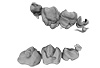
This contribution contains the 3D models described and figured in the following publication: Pujos F., Hautier L., Antoine P-O., Boivin M., Moison B, Salas-Gismondi R, Tejada J.V. , Varas-Malca R.M., Yans J., Marivaux L. (2025). Unexpected pampatheriid from the early Oligocene of Peruvian Amazonia: insights into the tropical differentiation of cingulate xenarthrans. Historical Biology.
Bradypus tridactylus UM-ZOOL-V69 View specimen
|
M3#1600Molariform and associated dentinal microstructure Type: "3D_surfaces"doi: 10.18563/m3.sf.1600 state:published |
Download 3D surface file |
Choloepus didactylus UM-ZOOL-V12 View specimen
|
M3#1601Molariform and associated dentinal microstructure Type: "3D_surfaces"doi: 10.18563/m3.sf.1601 state:published |
Download 3D surface file |
Dasypus mexicanus UM-ZOOL-2787 View specimen
|
M3#1602Molariform and associated dentinal microstructure Type: "3D_surfaces"doi: 10.18563/m3.sf.1602 state:published |
Download 3D surface file |
Tolypeutes matacus UM-ZOOL-2789 View specimen
|
M3#1603Molariform and associated dentinal microstructure Type: "3D_surfaces"doi: 10.18563/m3.sf.1603 state:published |
Download 3D surface file |
Euphractus sexcinctus UM-ZOOL-2790 View specimen
|
M3#1604Molariform and associated dentinal microstructure Type: "3D_surfaces"doi: 10.18563/m3.sf.1604 state:published |
Download 3D surface file |
Holmesina septrionalis UM-FLD-1 View specimen
|
M3#1605Molariform and associated dentinal microstructure Type: "3D_surfaces"doi: 10.18563/m3.sf.1605 state:published |
Download 3D surface file |
Megatherium sp. UM-TAR-1 View specimen
|
M3#1607Molariform and associated dentinal microstructure Type: "3D_surfaces"doi: 10.18563/m3.sf.1607 state:published |
Download 3D surface file |
Indet indet MUSM-3965 View specimen
|
M3#1606Molariform and associated dentinal microstructure Type: "3D_surfaces"doi: 10.18563/m3.sf.1606 state:published |
Download 3D surface file |
The present 3D Dataset contains the 3D models analyzed in: Hirose, A., Nakashima, T., Yamada, S., Uwabe, C., Kose, K., Takakuwa, T. 2012. Embryonic liver morphology and morphometry by magnetic resonance microscopic imaging. Anat Rec (Hoboken) 295, 51-59. doi: 10.1002/ar.21496
Homo sapiens KC-CS14LIV1387 View specimen
|
M3#64Human liver at Carnegie Stage (CS) 14 Type: "3D_surfaces"doi: 10.18563/m3.sf.64 state:published |
Download 3D surface file |
Homo sapiens KC-CS15LIV5074 View specimen
|
M3#65Human liver at Carnegie Stage (CS) 15 Type: "3D_surfaces"doi: 10.18563/m3.sf.65 state:published |
Download 3D surface file |
Homo sapiens KC-CS16LIV2578 View specimen
|
M3#66Human liver at Carnegie Stage (CS) 16 Type: "3D_surfaces"doi: 10.18563/m3.sf.66 state:published |
Download 3D surface file |
Homo sapiens KC-CS17LIV17832 View specimen
|
M3#67Human liver at Carnegie Stage (CS) 17 Type: "3D_surfaces"doi: 10.18563/m3.sf.67 state:published |
Download 3D surface file |
Homo sapiens KC-CS18LIV21124 View specimen
|
M3#68Human liver at Carnegie Stage (CS) 18 Type: "3D_surfaces"doi: 10.18563/m3.sf.68 state:published |
Download 3D surface file |
Homo sapiens KC-CS19LIV14353 View specimen

|
M3#69Human liver at Carnegie Stage (CS) 19 Type: "3D_surfaces"doi: 10.18563/m3.sf.69 state:published |
Download 3D surface file |
Homo sapiens KC-CS20LIV20701 View specimen
|
M3#70Human liver at Carnegie Stage (CS) 20 Type: "3D_surfaces"doi: 10.18563/m3.sf.70 state:published |
Download 3D surface file |
Homo sapiens KC-CS21LIV25858 View specimen
|
M3#71Human liver at Carnegie Stage (CS) 21 Type: "3D_surfaces"doi: 10.18563/m3.sf.71 state:published |
Download 3D surface file |
Homo sapiens KC-CS22LIV22226 View specimen
|
M3#72Human liver at Carnegie Stage (CS) 22 Type: "3D_surfaces"doi: 10.18563/m3.sf.72 state:published |
Download 3D surface file |
Homo sapiens KC-CS23LIV25704 View specimen
|
M3#73Human liver at Carnegie Stage (CS) 23 Type: "3D_surfaces"doi: 10.18563/m3.sf.73 state:published |
Download 3D surface file |
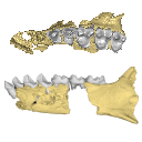
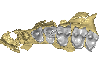
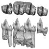
The present 3D Dataset contains the 3D models analyzed in Hendrickx, C., Gaetano, L. C., Choiniere, J., Mocke, H. and Abdala, F. in press. A new traversodontid cynodont with a peculiar postcanine dentition from the Middle/Late Triassic of Namibia and dental evolution in basal gomphodonts. Journal of Systematic Palaeontology.
Etjoia dentitransitus GSN F1591 View specimen
|
M3#557Surface model derived from µCT data of the holotype of Etjoia dentitransitus Type: "3D_surfaces"doi: 10.18563/m3.sf.557 state:published |
Download 3D surface file |
|
M3#558Photogrammetric 3D surface model of the postcanines of the Holotype of Etjoia dentitransitus Type: "3D_surfaces"doi: 10.18563/m3.sf.558 state:published |
Download 3D surface file |
|
M3#559Photogrammetric 3D surface model of the Holotype of Etjoia dentitransitus Type: "3D_surfaces"doi: 10.18563/m3.sf.559 state:published |
Download 3D surface file |
The present 3D Dataset contains the 3D models of the sacral vertebrae analyzed in “Sacral co-ossification in dinosaurs: The oldest record of fused sacral vertebrae in Dinosauria and the diversity of sacral co-ossification patterns in the group”.
Buriolestes schultzi CAPPA/UFSM 0035 View specimen

|
M3#705Sacral vertebrae of Buriolestes schultzi Type: "3D_surfaces"doi: 10.18563/m3.sf.705 state:published |
Download 3D surface file |
indet indet CAPPA/UFSM 0228 View specimen
|
M3#706Sacral vertebrae of a saurischian dinosaur indet. Type: "3D_surfaces"doi: 10.18563/m3.sf.706 state:published |
Download 3D surface file |
This contribution contains the 3D models described and figured in the following publication: Paulina-Carabajal, A. and Nieto, M. N. In press. Brief comment on the brain and inner ear of Giganotosaurus carolinii (Dinosauria: Theropoda) based on CT scans. Ameghiniana. https://doi.org/10.5710/AMGH.25.10.2019.3237
Giganotosaurus carolinii MUCPv-CH-1 View specimen
|
M3#504The current file contents 3D models of the braincase, brain, left and right inner ears Type: "3D_surfaces"doi: 10.18563/m3.sf.504 state:published |
Download 3D surface file |
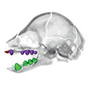
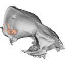
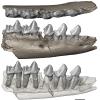
This contribution contains the 3D models described and figured in the following publication: Hautier L., Gomes Rodrigues H., Billet G., Asher R.J., 2016. The hidden teeth of sloths: evolutionary vestiges and the development of a simplified dentition. Scientific Reports. doi: 10.1038/srep27763
Bradypus variegatus ZMB 33812 View specimen
|
M3#110Three-dimensional reconstruction of the teeth, mandibles, maxillary and premaxillary bones Type: "3D_surfaces"doi: 10.18563/m3.sf.110 state:published |
Download 3D surface file |
Bradypus variegatus ZMB 41122 View specimen
|
M3#109Three-dimensional reconstruction of the teeth, mandibles, maxillary and premaxillary bones Type: "3D_surfaces"doi: 10.18563/m3.sf.109 state:published |
Download 3D surface file |
Bradypus variegatus MNHN-ZM-MO-1995-326A View specimen
|
M3#111Three-dimensional reconstruction of the teeth, mandibles, maxillary and premaxillary bones Type: "3D_surfaces"doi: 10.18563/m3.sf.111 state:published |
Download 3D surface file |
Bradypus variegatus MNHN-ZM-MO-1995-326B View specimen
|
M3#112Three-dimensional reconstruction of the teeth, mandibles, maxillary and premaxillary bones Type: "3D_surfaces"doi: 10.18563/m3.sf.112 state:published |
Download 3D surface file |
Bradypus sp. MNHN-ZM-MO-1902-325 View specimen
|
M3#113Three-dimensional reconstruction of the teeth, mandibles, maxillary, and premaxillary bones Type: "3D_surfaces"doi: 10.18563/m3.sf.113 state:published |
Download 3D surface file |
Bradypus sp. MNHN-ZM-MO-1995-327 View specimen
|
M3#114Three-dimensional reconstruction of the teeth, mandibles, maxillary and premaxillary bones Type: "3D_surfaces"doi: 10.18563/m3.sf.114 state:published |
Download 3D surface file |
Choloepus didactylus MNHN-ZM-MO-1882-625 View specimen
|
M3#115Three-dimensional reconstruction of the teeth, mandibles, maxillary and premaxillary bones Type: "3D_surfaces"doi: 10.18563/m3.sf.115 state:published |
Download 3D surface file |
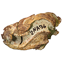
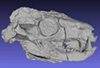
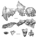
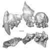
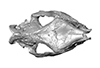
This contribution contains the 3D models described and figured in the following publication: Kassegne K. E., Mourlam M. J., Guinot G., Amoudji Y. Z., Martin J. E., Togbe K. A., Johnson A. K., Hautier L. 2021. First partial cranium of Togocetus from Kpogamé (Togo) and the protocetid diversity in the Togolese phosphate basin. Annales de Paléontologie, Issue 2, April–June 2021, 102488. https://doi.org/10.1016/j.annpal.2021.102488
Togocetus cf. traversei ULDG-KPO1 View specimen
|
M3#768The specimen consists of a partial cranium prepared out of a calcareous phosphate matrix. The partial cranium lacks the anterior part of the rostrum, the cranial roof, and most of the basicranium apart from the left zygomatic process of the squamosal. The maxilla, nasal, palatine, pterygoid, alisphenoid, and squamosal bones are preserved, as well as two incomplete dental rows described hereafter. Type: "3D_surfaces"doi: 10.18563/m3.sf.768 state:published |
Download 3D surface file |
|
M3#770µCT . Resolution: 0.3156mm. This scan can easily be opened with Fiji, MorphoDig, 3DSlicer, or any software that reads .MHD file format. Also, the .RAW file can be opened easily with other software such as Avizo/Amira when providing the correct dimensions (which are enclosed within the file name) Type: "3D_CT"doi: 10.18563/m3.sf.770 state:published |
Download CT data |
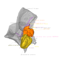
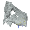
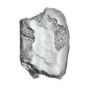
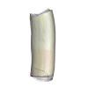
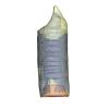
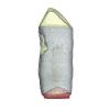
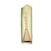
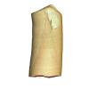
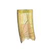
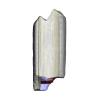
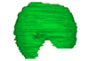
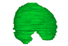
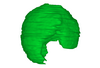
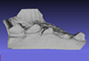
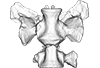
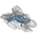
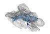
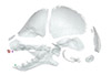
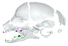
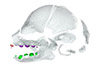
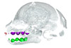
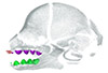
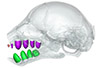
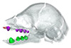
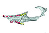
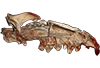
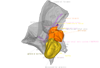
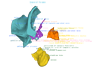
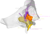
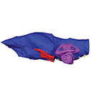
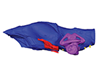
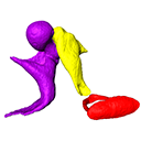
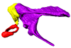
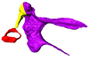
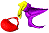
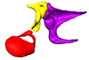
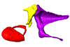
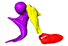
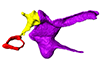
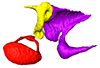
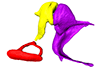
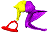
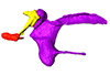
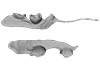
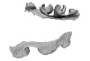
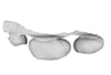
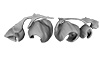
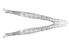
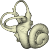
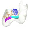
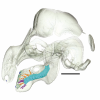
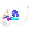
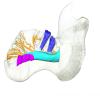
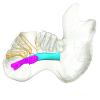
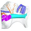
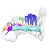
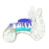
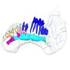
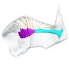
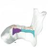
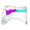
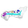
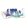
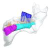
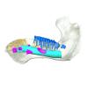
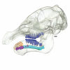
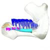
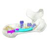
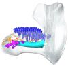
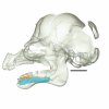
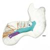
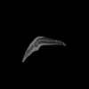
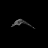
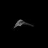
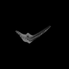
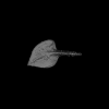
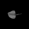
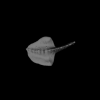
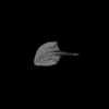
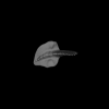
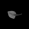
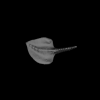
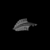
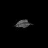
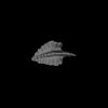
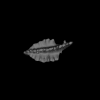
This project presents the osteological connexions of the petrosal bone of the extant Hippopotamidae Hippopotamus amphibius and Choeropsis liberiensis by a virtual osteological dissection of the ear region. The petrosal, the bulla, the sinuses and the major morphological features surrounding the petrosal bone are labelled, both in situ and in an exploded model presenting disassembly views. The directional underwater hearing mode of Hippopotamidae is discussed based on the new observations.
Choeropsis liberiensis UPPal-M09-5-005a View specimen
|
M3#1Labelled compact model of the right ear region of Choeropsis liberiensis (UPPal-M09-5-005a) Type: "3D_surfaces"doi: 10.18563/m3.sf1 state:published |
Download 3D surface file |
|
M3#2Labelled exploded model of the right ear region of Choeropsis liberiensis (UPPal-M09-5-005a) Type: "3D_surfaces"doi: 10.18563/m3.sf2 state:published |
Download 3D surface file |
Hippopotamus amphibius UM N179 View specimen
|
M3#3Labelled compact model of the right ear region of Hippopotamus amphibius (UM N 179) Type: "3D_surfaces"doi: 10.18563/m3.sf3 state:published |
Download 3D surface file |

|
M3#4Labelled exploded model of the right ear region of Hippopotamus amphibius (UM N 179) Type: "3D_surfaces"doi: 10.18563/m3.sf4 state:published |
Download 3D surface file |
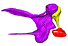
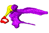
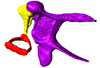
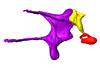
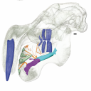
The present publication contains the µCT dataset and the 3D models analyzed in the following publication: Mautner, A.-K., A. E. Latimer, U. Fritz, and T. M. Scheyer. An updated description of the osteology of the pancake tortoise Malacochersus tornieri (Testudines: Testudinidae) with special focus on intraspecific variation. Journal of Morphology. https://doi.org/10.1002/jmor.20640
Malacochersus tornieri ZM 100.102 View specimen
|
M3#129Virtual brain and inner ear endocast of Malacochersus tornieri (ZM 100.102; Zoological Museum of The University of Zurich). This virtual model is accompanied by the 3D dataset. Blue, endocranium; red, blood vessels; purple, semicircular canals; yellow, cranial nerves. Type: "3D_surfaces"doi: 10.18563/m3.sf.129 state:published |
Download 3D surface file |
|
M3#1303D dataset of skull of Malacochersus tornieri (ZM 100.102) Type: "3D_CT"doi: 10.18563/m3.sf.130 state:published |
Download CT data |
Considerable morphological variations are found in the middle ear among mammals. Here I present a three-dimensional atlas of the middle ear ossicles of eulipotyphlan mammals. This group has radiated into various environments as terrestrial, aquatic, and subterranean habitats independently in multiple lineages. Therefore, eulipotyphlans are an ideal group to explore the form-function relationship of the middle ear ossicles. This comparative atlas of hedgehogs, true shrews, water shrews, mole shrews, true moles, and shrew moles encourages future studies of the middle ear morphology of this diverse group.
Erinaceus europaeus DK2331 View specimen
|
M3#151Left middle ear ossicles Type: "3D_surfaces"doi: 10.18563/m3.sf.151 state:published |
Download 3D surface file |
Anourosorex yamashinai SIK_yamashinai View specimen
|
M3#152Left middle ear ossicles Type: "3D_surfaces"doi: 10.18563/m3.sf.152 state:published |
Download 3D surface file |
Blarina brevicauda M8003 View specimen
|
M3#153Right middle ear ossicles Type: "3D_surfaces"doi: 10.18563/m3.sf.153 state:published |
Download 3D surface file |
Chimarrogale platycephala DK5481 View specimen
|
M3#162Left middle ear ossicles Type: "3D_surfaces"doi: 10.18563/m3.sf.162 state:published |
Download 3D surface file |
Suncus murinus DK1227 View specimen
|
M3#155Left middle ear ossicles Type: "3D_surfaces"doi: 10.18563/m3.sf.155 state:published |
Download 3D surface file |
Condylura cristata SIK0050 View specimen
|
M3#156Right middle ear ossicles Type: "3D_surfaces"doi: 10.18563/m3.sf.156 state:published |
Download 3D surface file |
Euroscaptor klossi SIK0673 View specimen
|
M3#163Left middle ear ossicles Type: "3D_surfaces"doi: 10.18563/m3.sf.163 state:published |
Download 3D surface file |
Euroscaptor malayana SIK_malayana View specimen
|
M3#164Left middle ear ossicles Type: "3D_surfaces"doi: 10.18563/m3.sf.164 state:published |
Download 3D surface file |
Mogera wogura DK2551 View specimen
|
M3#159Left middle ear ossicles Type: "3D_surfaces"doi: 10.18563/m3.sf.159 state:published |
Download 3D surface file |
Talpa altaica SIK_altaica View specimen
|
M3#161Right middle ear ossicles Type: "3D_surfaces"doi: 10.18563/m3.sf.161 state:published |
Download 3D surface file |
Urotrichus talpoides DK0887 View specimen
|
M3#165Left middle ear ossicles Type: "3D_surfaces"doi: 10.18563/m3.sf.165 state:published |
Download 3D surface file |
Oreoscaptor mizura DK6545 View specimen
|
M3#166Left middle ear ossicles Type: "3D_surfaces"doi: 10.18563/m3.sf.166 state:published |
Download 3D surface file |
Scalopus aquaticus SIK_aquaticus View specimen
|
M3#167Left middle ear ossicles Type: "3D_surfaces"doi: 10.18563/m3.sf.167 state:published |
Download 3D surface file |
Scapanus orarius SIK_orarius View specimen
|
M3#168Left middle ear ossicles Type: "3D_surfaces"doi: 10.18563/m3.sf.168 state:published |
Download 3D surface file |
Neurotrichus gibbsii SIK_gibbsii View specimen
|
M3#169Left middle ear ossicles Type: "3D_surfaces"doi: 10.18563/m3.sf.169 state:published |
Download 3D surface file |
The present 3D Dataset contains the 3D models analyzed in ”Morphological features of tooth development and replacement in the rabbit Oryctolagus cuniculus”, Archives of Oral Biology, https://doi.org/10.1016/j.archoralbio.2019.104576
Oryctogalus cuniculus E14 View specimen
|
M3#390Right cheek teeth, Left and right incisors at 14 dpf Type: "3D_surfaces"doi: 10.18563/m3.sf.390 state:published |
Download 3D surface file |
Oryctogalus cuniculus E16 View specimen
|
M3#391Left cheek teeth, Left and right incisors at 16 dpf Type: "3D_surfaces"doi: 10.18563/m3.sf.391 state:published |
Download 3D surface file |
Oryctogalus cuniculus E18 View specimen
|
M3#392Left cheek teeth and incisors at 18 dpf Type: "3D_surfaces"doi: 10.18563/m3.sf.392 state:published |
Download 3D surface file |
Oryctogalus cuniculus E20 View specimen
|
M3#393Left cheek teeth and incisors at 20 dpf Type: "3D_surfaces"doi: 10.18563/m3.sf.393 state:published |
Download 3D surface file |
Oryctogalus cuniculus E22 View specimen
|
M3#394Left lower cheek teeth and incisors, right upper cheek teeth and incisors at 22 dpf Type: "3D_surfaces"doi: 10.18563/m3.sf.394 state:published |
Download 3D surface file |
Oryctogalus cuniculus E24 View specimen
|
M3#395Left cheek teeth and incisors at 24 dpf Type: "3D_surfaces"doi: 10.18563/m3.sf.395 state:published |
Download 3D surface file |
Oryctogalus cuniculus E28 View specimen
|
M3#396Right cheek teeth and incisors at 28 dpf Type: "3D_surfaces"doi: 10.18563/m3.sf.396 state:published |
Download 3D surface file |
Oryctogalus cuniculus E26 View specimen
|
M3#397Right cheek teeth and incisors at 26 dpf Type: "3D_surfaces"doi: 10.18563/m3.sf.397 state:published |
Download 3D surface file |
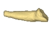
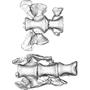
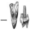
This note presents the 3D model of the hemi-mandible UM-PAT 159 of the MP7 Diacodexis species D. cf. gigasei and 3D models corresponding to the restoration of the ascending ramus, broken on the original specimen, and to a restoration of a complete mandible based on the preserved left hemi-mandible.
Diacodexis cf. gigasei UMPAT159 View specimen
|
M3#3153D models of UM PAT 159 after the restoration of the ascending ramus Type: "3D_surfaces"doi: 10.18563/m3.sf.315 state:published |
Download 3D surface file |
|
M3#316restoration of a complete mandible based on the preserved left hemi-mandible UM PAT 159 Type: "3D_surfaces"doi: 10.18563/m3.sf.316 state:published |
Download 3D surface file |
|
M3#3173D model of the hemi-mandible UM PAT 159 Type: "3D_surfaces"doi: 10.18563/m3.sf.317 state:published |
Download 3D surface file |
The present 3D Dataset contains 3D models of the cranium surface and of the bony labyrinth endocast of the stem bat Vielasia sigei. They are used by (Hand et al., 2023) to explore the phylogenetic position of this species, to infer its laryngeal echolocating capabilities, and to eventually discuss chiropteran evolution before the crown clade diversification.
Vielasia sigei UM VIE-250 View specimen
|
M3#1269External surface of the cranium Type: "3D_surfaces"doi: 10.18563/m3.sf.1269 state:published |
Download 3D surface file |
|
M3#1270Virtual endocast of the right bony labyrinth Type: "3D_surfaces"doi: 10.18563/m3.sf.1270 state:published |
Download 3D surface file |
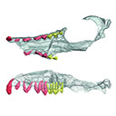
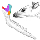
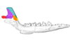
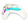
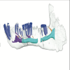
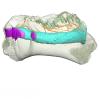
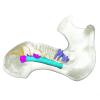
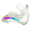
This contribution contains the 3D models described and figured in the following publication: Hautier L, Gomes Rodrigues H, Ferreira-Cardoso S, Emerling CA, Porcher M-L, Asher R, Portela Miguez R, Delsuc F. 2023. From teeth to pad: tooth loss and development of keratinous structures in sirenians. Proceedings of the Royal Society B. https://doi.org/10.1098/rspb.2023.1932
Dugong dugon 2005.51 View specimen

|
M3#1275Internal mandibular morphology. Orange = dorsal canaliculi; purple = mental branches; cyan = mandibular canal; dark blue = teeth; green = tooth alveoli. Type: "3D_surfaces"doi: 10.18563/m3.sf.1275 state:published |
Download 3D surface file |
Dugong dugon 2023.66 View specimen
|
M3#1274Internal mandibular morphology. Orange = dorsal canaliculi; purple = mental branches; cyan = mandibular canal; dark blue = teeth; green = tooth alveoli. Type: "3D_surfaces"doi: 10.18563/m3.sf.1274 state:published |
Download 3D surface file |
Dugong dugon 5386 View specimen
|
M3#1276Internal mandibular morphology. Orange = dorsal canaliculi; purple = mental branches; cyan = mandibular canal; dark blue = teeth; green = tooth alveoli. Type: "3D_surfaces"doi: 10.18563/m3.sf.1276 state:published |
Download 3D surface file |
Dugong dugon 1848.8.29.7/GERM 1027g View specimen
|
M3#1277Internal mandibular morphology. Orange = dorsal canaliculi; purple = mental branches; cyan = mandibular canal; dark blue = teeth. Type: "3D_surfaces"doi: 10.18563/m3.sf.1277 state:published |
Download 3D surface file |
Dugong dugon 1991.413 View specimen
|
M3#1278Internal mandibular morphology. Orange = dorsal canaliculi; purple = mental branches; cyan = mandibular canal; dark blue = teeth. Type: "3D_surfaces"doi: 10.18563/m3.sf.1278 state:published |
Download 3D surface file |
Dugong dugon 1991.427 View specimen
|
M3#1279Internal mandibular morphology. Orange = dorsal canaliculi; purple = mental branches; cyan = mandibular canal; dark blue = teeth. Type: "3D_surfaces"doi: 10.18563/m3.sf.1279 state:published |
Download 3D surface file |
Dugong dugon 2017-3-9 View specimen
|
M3#1280Internal mandibular morphology. Orange = dorsal canaliculi; purple = mental branches; cyan = mandibular canal; dark blue = teeth. Type: "3D_surfaces"doi: 10.18563/m3.sf.1280 state:published |
Download 3D surface file |
Eosiren lybica 1913-22 View specimen
|
M3#1281Internal mandibular morphology. Orange = dorsal canaliculi; purple = mental branches; cyan = mandibular canal; dark blue = teeth; green = tooth alveoli. Type: "3D_surfaces"doi: 10.18563/m3.sf.1281 state:published |
Download 3D surface file |
Halitherium taulannense RGHP C001 View specimen
|
M3#1282Internal mandibular morphology. Cyan = mandibular canal; dark blue = teeth. Type: "3D_surfaces"doi: 10.18563/m3.sf.1282 state:published |
Download 3D surface file |
Halitherium taulannense RGHP C009 View specimen
|
M3#1283Internal mandibular morphology. Orange = dorsal canaliculi; purple = mental branches; cyan = mandibular canal; dark blue = teeth; green = tooth alveoli. Type: "3D_surfaces"doi: 10.18563/m3.sf.1283 state:published |
Download 3D surface file |
Hydrodamalis gigas 1947.10.21.1 View specimen
|
M3#1284Internal mandibular morphology. Orange = dorsal canaliculi; purple = mental branches; cyan = mandibular canal Type: "3D_surfaces"doi: 10.18563/m3.sf.1284 state:published |
Download 3D surface file |
Hydrodamalis gigas C1021 View specimen
|
M3#1285Anterior part of the mandible Type: "3D_surfaces"doi: 10.18563/m3.sf.1285 state:published |
Download 3D surface file |

|
M3#1286Posterior part of the mandible Type: "3D_surfaces"doi: 10.18563/m3.sf.1286 state:published |
Download 3D surface file |
Hydrodamalis gigas 2023.67 View specimen
|
M3#1287Internal mandibular morphology. Orange = dorsal canaliculi; purple = mental branches; cyan = mandibular canal Type: "3D_surfaces"doi: 10.18563/m3.sf.1287 state:published |
Download 3D surface file |
Libysiren sickenbergi M.82429 View specimen
|
M3#1288Internal mandibular morphology. Orange = dorsal canaliculi; purple = mental branches; cyan = mandibular canal; dark blue = teeth; green = tooth alveoli. Type: "3D_surfaces"doi: 10.18563/m3.sf.1288 state:published |
Download 3D surface file |
Libysiren sickenbergi M.45675 View specimen
|
M3#1289Internal mandibular morphology. Orange = dorsal canaliculi; purple = mental branches; cyan = mandibular canal; dark blue = teeth; green = tooth alveoli. Type: "3D_surfaces"doi: 10.18563/m3.sf.1289 state:published |
Download 3D surface file |
Prorastomus sirenoides OR.448976 View specimen
|
M3#1290Internal morphology of the left mandible. Orange = dorsal canaliculi; purple = mental branches; cyan = mandibular canal; dark blue = teeth; green = tooth alveoli. Type: "3D_surfaces"doi: 10.18563/m3.sf.1290 state:published |
Download 3D surface file |
|
M3#1304Internal morphology of the right mandible. Orange = dorsal canaliculi; purple = mental branches; cyan = mandibular canal; dark blue = teeth. Type: "3D_surfaces"doi: 10.18563/m3.sf.1304 state:published |
Download 3D surface file |
Ribodon limbatus M.7073 View specimen
|
M3#1292Internal mandibular morphology. Orange = dorsal canaliculi; purple = mental branches; cyan = mandibular canal; dark blue = teeth; green = tooth alveoli. Type: "3D_surfaces"doi: 10.18563/m3.sf.1292 state:published |
Download 3D surface file |
Rytiodus capgrandi PAL2017-8-1 View specimen
|
M3#1293Internal mandibular morphology. Orange = dorsal canaliculi; purple = mental branches; cyan = mandibular canal; dark blue = teeth; green = tooth alveoli. Type: "3D_surfaces"doi: 10.18563/m3.sf.1293 state:published |
Download 3D surface file |
Trichechus inunguis 1868.12.19.2 View specimen
|
M3#1294Internal mandibular morphology. Orange = dorsal canaliculi; purple = mental branches; cyan = mandibular canal; dark blue = teeth; green = tooth alveoli. Type: "3D_surfaces"doi: 10.18563/m3.sf.1294 state:published |
Download 3D surface file |
Trichechus manatus 1843.3.10.12 View specimen
|
M3#1295Internal mandibular morphology. Orange = dorsal canaliculi; purple = mental branches; cyan = mandibular canal; dark blue = teeth. Type: "3D_surfaces"doi: 10.18563/m3.sf.1295 state:published |
Download 3D surface file |
Trichechus manatus 1864.6.5.1 View specimen
|
M3#1296Internal mandibular morphology. Orange = dorsal canaliculi; purple = mental branches; cyan = mandibular canal; dark blue = teeth; green = tooth alveoli. Type: "3D_surfaces"doi: 10.18563/m3.sf.1296 state:published |
Download 3D surface file |
Trichechus manatus 1950.1.23.1 View specimen
|
M3#1297Internal mandibular morphology. Orange = dorsal canaliculi; purple = mental branches; cyan = mandibular canal; dark blue = teeth; green = tooth alveoli. Type: "3D_surfaces"doi: 10.18563/m3.sf.1297 state:published |
Download 3D surface file |
Trichechus senegalensis 1885.6.30.2 View specimen
|
M3#1298Internal mandibular morphology. Orange = dorsal canaliculi; purple = mental branches; cyan = mandibular canal; dark blue = teeth; green = tooth alveoli. Type: "3D_surfaces"doi: 10.18563/m3.sf.1298 state:published |
Download 3D surface file |
Trichechus senegalensis 1894.7.25.8 View specimen
|
M3#1299Internal mandibular morphology. Orange = dorsal canaliculi; purple = mental branches; cyan = mandibular canal; dark blue = teeth. Type: "3D_surfaces"doi: 10.18563/m3.sf.1299 state:published |
Download 3D surface file |
Trichechus senegalensis V97 View specimen
|
M3#1302Mandibular internal morphology. Orange = dorsal canaliculi; purple = mental branches; cyan = mandibular canal; dark blue = teeth. Type: "3D_surfaces"doi: 10.18563/m3.sf.1302 state:published |
Download 3D surface file |
Trichechus sp. 65.4.28.9 View specimen
|
M3#1300Internal mandibular morphology. Orange = dorsal canaliculi; purple = mental branches; cyan = mandibular canal; dark blue = teeth; green = tooth alveoli. Type: "3D_surfaces"doi: 10.18563/m3.sf.1300 state:published |
Download 3D surface file |
Dugong dugon 1946.8.6.2 View specimen
|
M3#1301Mandibular internal morphology. Orange = dorsal canaliculi; purple = mental branches; cyan = mandibular canal; dark blue = teeth. Type: "3D_surfaces"doi: 10.18563/m3.sf.1301 state:published |
Download 3D surface file |
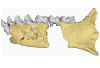
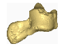
This contribution contains the 3D models of the fossil remains (maxilla, dentary, and talus) attributed to Djebelemur martinezi, a ca. 50 Ma primate from Tunisia (Djebel Chambi), described and figured in the following publication: Marivaux et al. (2013), Djebelemur, a tiny pre-tooth-combed primate from the Eocene of Tunisia: a glimpse into the origin of crown strepsirhines. PLoS ONE. https://doi.org/10.1371/journal.pone.0080778
Djebelemur martinezi CBI-1-544 View specimen
|
M3#365CBI-1-544, left maxilla preserving P3-M3 and alveoli for P2 and C1 Type: "3D_surfaces"doi: 10.18563/m3.sf.365 state:published |
Download 3D surface file |
Djebelemur martinezi CBI-1-567 View specimen
|
M3#363Isolated left upper P4 Type: "3D_surfaces"doi: 10.18563/m3.sf.363 state:published |
Download 3D surface file |
Djebelemur martinezi CBI-1-565-577-587-580 View specimen
|
M3#366- CBI-1-565, a damaged right mandible, which consists of three isolated pieces found together and reassembled here: the anterior part of the dentary bears the p3 and m1, and alveoli for p4, p2 and c, while the posterior part preserves m3 and a portion of the ascending ramus; the m2 was found isolated but in the same small calcareous block treated by acid processing. - CBI-1-577, isolated right lower p4. - CBI-1-587, isolated left lower p2 (reversed). - CBI-1-580, isolated left lower canine (reversed). Type: "3D_surfaces"doi: 10.18563/m3.sf.366 state:published |
Download 3D surface file |
Djebelemur martinezi CBI-1-545 View specimen
|
M3#364Right Talus Type: "3D_surfaces"doi: 10.18563/m3.sf.364 state:published |
Download 3D surface file |
This contribution contains the three-dimensional models of the most complete and/or informative fossil materials attributed to Peradectes crocheti Gernelle, 2024, the earliest peradectid metatherian species of Europe, from its type locality (Palette, Provence, ~55 Ma). These specimens were analyzed and discussed in: Gernelle et al. (2024), Taxonomy and evolutionary history of peradectids (Metatheria): new data from the early Eocene of France. https://doi.org/10.1007/s10914-024-09724-5
Peradectes crocheti MHN.AIX.PV.2018.26.14 View specimen
|
M3#14993D surface model of MHN.AIX.PV.2018.26.14, fragmentary left maxilla with C-P1, anterior root of P2, and M1-M3 Type: "3D_surfaces"doi: 10.18563/m3.sf.1499 state:published |
Download 3D surface file |
Peradectes crocheti MHN.AIX.PV.2017.6.6 View specimen
|
M3#15003D surface model of MHN.AIX.PV.2017.6.6, left P2 Type: "3D_surfaces"doi: 10.18563/m3.sf.1500 state:published |
Download 3D surface file |
Peradectes crocheti MHN.AIX.PV.2017.6.7 View specimen
|
M3#15013D surface model of MHN.AIX.PV.2017.6.7, left M3 Type: "3D_surfaces"doi: 10.18563/m3.sf.1501 state:published |
Download 3D surface file |
Peradectes crocheti MHN.AIX.PV.2017.6.8 View specimen
|
M3#15023D surface model of MHN.AIX.PV.2017.6.8, right hemi-mandible fragment with canine alveolus, posterior root of p1, partial p2, p3, partial m1, and m2-m3 Type: "3D_surfaces"doi: 10.18563/m3.sf.1502 state:published |
Download 3D surface file |
Peradectes crocheti MHN.AIX.PV.2017.6.9 View specimen
|
M3#15033D surface model of MHN.AIX.PV.2017.6.9, leftm1-m4 row with fragments of dentary Type: "3D_surfaces"doi: 10.18563/m3.sf.1503 state:published |
Download 3D surface file |
Peradectes crocheti MHN.AIX.PV.2017.6.14 View specimen
|
M3#15043D surface model of MHN.AIX.PV.2017.6.14, right astragalus Type: "3D_surfaces"doi: 10.18563/m3.sf.1504 state:published |
Download 3D surface file |
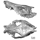
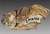
This contribution provides for the first time the 3D model of the type specimen of Molassitherium delemontense (Mammalia, Rhinocerotidae) described in the following publication: Becker et al. (2013), Journal of Systematic Palaeontology, Vol. 11, Issue 8, 947–972, https://doi.org/10.1080/14772019.2012.699007. Conservation issues of the specimen and solutions using 3D model and 3D prints are detailed.
Molassitherium delemontense MJSN POI007–245 View specimen
|
M3#384Skull of Molassitherium delemontense Becker and Antoine, 2013 (in Becker et al. 2013): holotype Type: "3D_surfaces"doi: 10.18563/m3.sf.384 state:published |
Download 3D surface file |
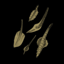
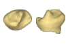
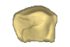
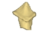
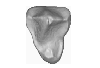
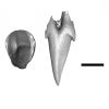
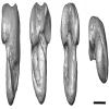
The present 3D Dataset contains the 3D models analyzed in Assemat et al. 2023: Shape diversity in conodont elements, a quantitative study using 3D topography. Marine Micropaleontology 184. https://doi.org/10.1016/j.marmicro.2023.102292
P1 elements represent dental components of the conodont apparatus that perform the final stage of food processing before ingestion. Consequently, quantifying the shape of P1 elements across the topographic indices of different conodont species becomes crucial for deciphering the diversity in feeding behavior within this group.
Bispathodus aculeatus UM CTB 082 View specimen
|
M3#1404P element Type: "3D_surfaces"doi: 10.18563/m3.sf.1404 state:published |
Download 3D surface file |
Bispathodus aculeatus UM CTB 083 View specimen

|
M3#1405P element Type: "3D_surfaces"doi: 10.18563/m3.sf.1405 state:published |
Download 3D surface file |
Bispathodus aculeatus UM CTB 086 View specimen
|
M3#1406P element Type: "3D_surfaces"doi: 10.18563/m3.sf.1406 state:published |
Download 3D surface file |
Bispathodus ultimus UM CTB 088 View specimen
|
M3#1407P element Type: "3D_surfaces"doi: 10.18563/m3.sf.1407 state:published |
Download 3D surface file |
Bispathodus aculeatus UM CTB 089 View specimen
|
M3#1408P element Type: "3D_surfaces"doi: 10.18563/m3.sf.1408 state:published |
Download 3D surface file |
Bispathodus costatus UM CTB 090 View specimen
|
M3#1409P element Type: "3D_surfaces"doi: 10.18563/m3.sf.1409 state:published |
Download 3D surface file |
Bispathodus ultimus UM CTB 092 View specimen
|
M3#1410P element Type: "3D_surfaces"doi: 10.18563/m3.sf.1410 state:published |
Download 3D surface file |
Bispathodus costatus UM CTB 093 View specimen
|
M3#1411P element Type: "3D_surfaces"doi: 10.18563/m3.sf.1411 state:published |
Download 3D surface file |
Bispathodus spinulicostatus UM CTB 094 View specimen
|
M3#1412P element Type: "3D_surfaces"doi: 10.18563/m3.sf.1412 state:published |
Download 3D surface file |
Bispathodus aculeatus UM CTB 096 View specimen

|
M3#1413P element Type: "3D_surfaces"doi: 10.18563/m3.sf.1413 state:published |
Download 3D surface file |
Bispathodus ultimus UM CTB 098 View specimen
|
M3#1414P element Type: "3D_surfaces"doi: 10.18563/m3.sf.1414 state:published |
Download 3D surface file |
Bispathodus costatus UM CTB 060 View specimen
|
M3#1415P element Type: "3D_surfaces"doi: 10.18563/m3.sf.1415 state:published |
Download 3D surface file |
Bispathodus spinulicostatus UM CTB 073 View specimen
|
M3#1416P element Type: "3D_surfaces"doi: 10.18563/m3.sf.1416 state:published |
Download 3D surface file |
Branmehla suprema UM CTB 049 View specimen

|
M3#1417P element Type: "3D_surfaces"doi: 10.18563/m3.sf.1417 state:published |
Download 3D surface file |
Branmehla inornata UM CTB 100 View specimen
|
M3#1418P element Type: "3D_surfaces"doi: 10.18563/m3.sf.1418 state:published |
Download 3D surface file |
Bispathodus stabilis (morphe 1) UM CTB 101 View specimen

|
M3#1419P element Type: "3D_surfaces"doi: 10.18563/m3.sf.1419 state:published |
Download 3D surface file |
Branmehla suprema UM CTB 102 View specimen

|
M3#1420P element Type: "3D_surfaces"doi: 10.18563/m3.sf.1420 state:published |
Download 3D surface file |
Branmehla suprema UM CTB 103 View specimen
|
M3#1421P element Type: "3D_surfaces"doi: 10.18563/m3.sf.1421 state:published |
Download 3D surface file |
Branmehla suprema UM CTB 104 View specimen

|
M3#1422P element Type: "3D_surfaces"doi: 10.18563/m3.sf.1422 state:published |
Download 3D surface file |
Branmehla suprema UM CTB 105 View specimen

|
M3#1423P element Type: "3D_surfaces"doi: 10.18563/m3.sf.1423 state:published |
Download 3D surface file |
Branmehla suprema UM CTB 106 View specimen
|
M3#1424P element Type: "3D_surfaces"doi: 10.18563/m3.sf.1424 state:published |
Download 3D surface file |
Branmehla suprema UM CTB 072 View specimen

|
M3#1425P element Type: "3D_surfaces"doi: 10.18563/m3.sf.1425 state:published |
Download 3D surface file |
Branmehla suprema UM CTB 107 View specimen
|
M3#1426P element Type: "3D_surfaces"doi: 10.18563/m3.sf.1426 state:published |
Download 3D surface file |
Branmehla suprema UM CTB 108 View specimen

|
M3#1427P element Type: "3D_surfaces"doi: 10.18563/m3.sf.1427 state:published |
Download 3D surface file |
Branmehla suprema UM CTB 109 View specimen
|
M3#1428P element Type: "3D_surfaces"doi: 10.18563/m3.sf.1428 state:published |
Download 3D surface file |
Bispathodus stabilis (morphe 1) UM CTB 110 View specimen

|
M3#1429P element Type: "3D_surfaces"doi: 10.18563/m3.sf.1429 state:published |
Download 3D surface file |
Palmatolepis gracilis UM CTB 112 View specimen

|
M3#1430P element Type: "3D_surfaces"doi: 10.18563/m3.sf.1430 state:published |
Download 3D surface file |
Palmatolepis gracilis UM CTB 061 View specimen
|
M3#1431P element Type: "3D_surfaces"doi: 10.18563/m3.sf.1431 state:published |
Download 3D surface file |
Palmatolepis gracilis UM CTB 115 View specimen
|
M3#1432P element Type: "3D_surfaces"doi: 10.18563/m3.sf.1432 state:published |
Download 3D surface file |
Palmatolepis gracilis UM CTB 116 View specimen

|
M3#1433P element Type: "3D_surfaces"doi: 10.18563/m3.sf.1433 state:published |
Download 3D surface file |
Palmatolepis gracilis UM CTB 117 View specimen

|
M3#1434P element Type: "3D_surfaces"doi: 10.18563/m3.sf.1434 state:published |
Download 3D surface file |
Palmatolepis gracilis UM CTB 062 View specimen

|
M3#1435P element Type: "3D_surfaces"doi: 10.18563/m3.sf.1435 state:published |
Download 3D surface file |
Palmatolepis gracilis UM CTB 118 View specimen
|
M3#1436P element Type: "3D_surfaces"doi: 10.18563/m3.sf.1436 state:published |
Download 3D surface file |
Palmatolepis gracilis UM CTB 119 View specimen
|
M3#1437P element Type: "3D_surfaces"doi: 10.18563/m3.sf.1437 state:published |
Download 3D surface file |
Palmatolepis gracilis UM CTB 120 View specimen
|
M3#1438P element Type: "3D_surfaces"doi: 10.18563/m3.sf.1438 state:published |
Download 3D surface file |
Polygnathus communis UM CTB 075 View specimen
|
M3#1439P element Type: "3D_surfaces"doi: 10.18563/m3.sf.1439 state:published |
Download 3D surface file |
Polygnathus communis UM CTB 121 View specimen
|
M3#1440P element Type: "3D_surfaces"doi: 10.18563/m3.sf.1440 state:published |
Download 3D surface file |
Polygnathus communis UM CTB 122 View specimen

|
M3#1441P element Type: "3D_surfaces"doi: 10.18563/m3.sf.1441 state:published |
Download 3D surface file |
Polygnathus communis UM CTB 123 View specimen
|
M3#1442P element Type: "3D_surfaces"doi: 10.18563/m3.sf.1442 state:published |
Download 3D surface file |
Polygnathus communis UM CTB 125 View specimen
|
M3#1443P element Type: "3D_surfaces"doi: 10.18563/m3.sf.1443 state:published |
Download 3D surface file |
Polygnathus communis UM CTB 126 View specimen
|
M3#1444P element Type: "3D_surfaces"doi: 10.18563/m3.sf.1444 state:published |
Download 3D surface file |
Polygnathus communis UM CTB 128 View specimen
|
M3#1445P element Type: "3D_surfaces"doi: 10.18563/m3.sf.1445 state:published |
Download 3D surface file |
Polygnathus communis UM CTB 130 View specimen
|
M3#1446P element Type: "3D_surfaces"doi: 10.18563/m3.sf.1446 state:published |
Download 3D surface file |
Polygnathus communis UM CTB 131 View specimen
|
M3#1447P element Type: "3D_surfaces"doi: 10.18563/m3.sf.1447 state:published |
Download 3D surface file |
Polygnathus communis UM CTB 132 View specimen
|
M3#1448P element Type: "3D_surfaces"doi: 10.18563/m3.sf.1448 state:published |
Download 3D surface file |
Polygnathus communis UM CTB 133 View specimen
|
M3#1449P element Type: "3D_surfaces"doi: 10.18563/m3.sf.1449 state:published |
Download 3D surface file |
Polygnathus symmetricus UM CTB 139 View specimen
|
M3#1450P element Type: "3D_surfaces"doi: 10.18563/m3.sf.1450 state:published |
Download 3D surface file |
Polygnathus symmetricus UM CTB 140 View specimen
|
M3#1451P element Type: "3D_surfaces"doi: 10.18563/m3.sf.1451 state:published |
Download 3D surface file |
Polygnathus symmetricus UM CTB 141 View specimen
|
M3#1452P element Type: "3D_surfaces"doi: 10.18563/m3.sf.1452 state:published |
Download 3D surface file |
Polygnathus symmetricus UM CTB 142 View specimen
|
M3#1453P element Type: "3D_surfaces"doi: 10.18563/m3.sf.1453 state:published |
Download 3D surface file |


