Explodable 3D Dog Skull for Veterinary Education
3D models of Ocnotherium skull
3D models of Kalakocetus, the earliest Cetacea
3D GM dataset of bird skeletal variation
Skeletal embryonic development in the catshark
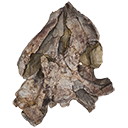
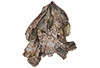
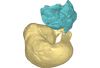
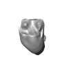
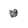
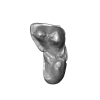
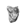
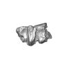
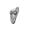
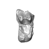
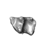
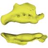
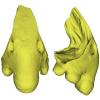
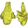
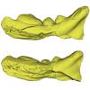
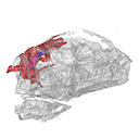
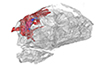
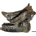
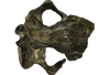
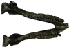
Bony connexions of the petrosal bone of extant hippos
bony labyrinth (14) , inner ear (11) , geometric morphometrics (10) , CT-scan (10) , Eocene (10) , Micro-CT (9) , Miocene (8)
Lionel Hautier (24) , Maëva Judith Orliac (23) , Laurent Marivaux (18) , Renaud Lebrun (14) , Rodolphe Tabuce (14) , Pierre-Olivier Antoine (13) , Bastien Mennecart (13)
|
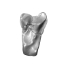
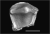
3D model related to the publication: A new species of the large-headed coastal marine turtle Solnhofia (Testudinata, Thalassochelydia) from the Late Jurassic of NW SwitzerlandJérémy Anquetin
Published online: 16/09/2020 |
|
M3#536Textured 3D surface model of the holotype cranium of the Late Jurassic turtle Solnhofia brachyrhyncha Type: "3D_surfaces"doi: 10.18563/m3.sf.536 state:published |
Download 3D surface file |
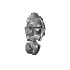
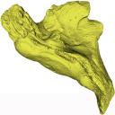
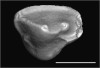
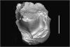
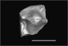
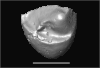
The present 3D Dataset contains the 3D model analyzed in the following publication: Paulina-Carabajal, A., Sterli, J., Werneburg, I., 2019. The endocranial anatomy of the stem turtle Naomichelys speciosa from the Early Cretaceous of North America. Acta Palaeontologica Polonica, https://doi.org/10.4202/app.00606.2019
Naomichelys speciosa FMNH PR273 View specimen
|
M3#428FMNH_PR273_1 - Naomichlys speciosa - skull Type: "3D_surfaces"doi: 10.18563/m3.sf.428 state:published |
Download 3D surface file |
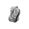
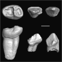
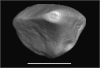
This contribution contains the 3D model of the holotype of Simplomys hugi, the new dormouse species from the locality of Glovelier described and figured in the following publication: New data on the Miocene dormouse Simplomys García-Paredes, 2009 from the peri-alpin basins of Switzerland and Germany: palaeodiversity of a rare genus in Central Europe. https://doi.org/10.1007/s12549-018-0339-y
Simplomys hugi MJSN-GLM017-0001 View specimen
|
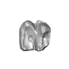
M3#385the left maxilla with four teeth ( DP4, P4, M1 and M2) Type: "3D_surfaces"doi: 10.18563/m3.sf.385 state:published |
Download 3D surface file |
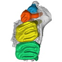
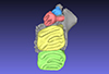
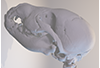
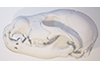
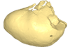
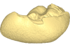
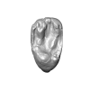
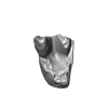
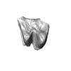
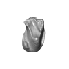
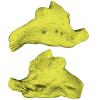
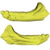
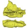
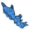
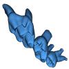
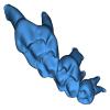
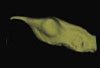
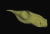
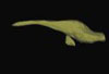
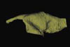
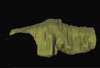
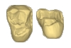
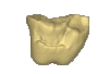
This contribution comprises the 3D models of three wolf pup skulls, which were used for the publication by Geiger et al. 2017 on Neomorphosis and heterochrony of skull shape in dog domestication.
Canis lupus CLL2 View specimen
|
M3#3123d model of a wolf pup skull Type: "3D_surfaces"doi: 10.18563/m3.sf.312 state:published |
Download 3D surface file |
Canis lupus CLL4 View specimen
|
M3#3133d model of a wolf pup skull Type: "3D_surfaces"doi: 10.18563/m3.sf.313 state:published |
Download 3D surface file |
Canis lupus CLL5 View specimen
|
M3#3143d model of a wolf pup skull Type: "3D_surfaces"doi: 10.18563/m3.sf.314 state:published |
Download 3D surface file |
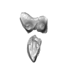
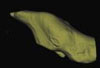
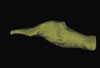
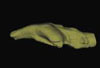
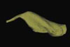
This contribution contains the 3D models described and figured in the following publication: Mourlam, M., Orliac, M. J. (2017), Protocetid (Cetacea, Artiodactyla) bullae and petrosals from the Middle Eocene locality of Kpogamé, Togo: new insights into the early history of cetacean hearing. Journal of Systematic Palaeontology https://doi.org/10.1080/14772019.2017.1328378
?Carolinacetus indet. UM KPG-M 164 View specimen
|
M3#132left petrosal of ?Carolinacetus sp. from the locality of Kpogamé, Togo Type: "3D_surfaces"doi: 10.18563/m3.sf.132 state:published |
Download 3D surface file |
indet. indet. UM KPG-M 73 View specimen

|
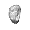
M3#133labelled surface of the left petrosal Type: "3D_surfaces"doi: 10.18563/m3.sf.133 state:published |
Download 3D surface file |
|
M3#134left bullaof Protocetidae indeterminate from Kpogamé, Togo Type: "3D_surfaces"doi: 10.18563/m3.sf.134 state:published |
Download 3D surface file |
|
M3#135petrotympanic complex of Protocetidae indeterminate from Kpogamé, Togo Type: "3D_surfaces"doi: 10.18563/m3.sf.135 state:published |
Download 3D surface file |
?Carolinacetus indet. UM KPG-M 33 View specimen
|
M3#136left auditory bulla of a juvenile specimen of ?Carolinacetus sp. from Kpogamé, Togo Type: "3D_surfaces"doi: 10.18563/m3.sf.136 state:published |
Download 3D surface file |
Togocetus traversei UM KPG-M 80 View specimen

|
M3#137fragmentary right auditory bulla of Togocetus traversei from Kpogamé, Togo Type: "3D_surfaces"doi: 10.18563/m3.sf.137 state:published |
Download 3D surface file |
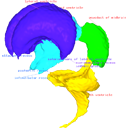
This contribution contains the 3D models described and figured in the following publication: Shiraishi N et al. Morphology and morphometry of the human embryonic brain: A three-dimensional analysis NeuroImage 115, 2015, 96-103, DOI: 10.1016/j.neuroimage.2015.04.044.
Homo sapiens KC-CS13BRN50455 View specimen

|
M3#24Computationally reconstructed cerebral parenchyma and ventricle of the human embryo at Carnegie Stage 13. Type: "3D_surfaces"doi: 10.18563/m3.sf24 state:published |
Download 3D surface file |
Homo sapiens KC-CS14BRN18834 View specimen

|
M3#25Computationally reconstructed cerebral parenchyma and ventricle of the human embryo at Carnegie Stage 14. Type: "3D_surfaces"doi: 10.18563/m3.sf25 state:published |
Download 3D surface file |
Homo sapiens KC-CS15BRN19975 View specimen

|
M3#26Computationally reconstructed cerebral parenchyma and ventricle of the human embryo at Carnegie Stage 15. Type: "3D_surfaces"doi: 10.18563/m3.sf26 state:published |
Download 3D surface file |
Homo sapiens KC-CS16BRN7870 View specimen
|
M3#27Computationally reconstructed cerebral parenchyma and ventricle of the human embryo at Carnegie Stage 16. Type: "3D_surfaces"doi: 10.18563/m3.sf27 state:published |
Download 3D surface file |
Homo sapiens KC-CS17BRN26702 View specimen

|
M3#28Computationally reconstructed cerebral parenchyma and ventricle of the human embryo at Carnegie Stage 17. Type: "3D_surfaces"doi: 10.18563/m3.sf28 state:published |
Download 3D surface file |
Homo sapiens KC-CS18BRN25914 View specimen

|
M3#29Computationally reconstructed cerebral parenchyma and ventricle of the human embryo at Carnegie Stage 18. Type: "3D_surfaces"doi: 10.18563/m3.sf29 state:published |
Download 3D surface file |
Homo sapiens KC-CS19BRN16508 View specimen

|
M3#30Computationally reconstructed cerebral parenchyma and ventricle of the human embryo at Carnegie Stage 19. Type: "3D_surfaces"doi: 10.18563/m3.sf30 state:published |
Download 3D surface file |
Homo sapiens KC-CS20BRN26581 View specimen

|
M3#31Computationally reconstructed cerebral parenchyma and ventricle of the human embryo at Carnegie Stage 20. Type: "3D_surfaces"doi: 10.18563/m3.sf31 state:published |
Download 3D surface file |
Homo sapiens KC-CS21BRN33434 View specimen

|
M3#32Computationally reconstructed cerebral parenchyma and ventricle of the human embryo at Carnegie Stage 21. Type: "3D_surfaces"doi: 10.18563/m3.sf32 state:published |
Download 3D surface file |
Homo sapiens KC-CS22BRN27960 View specimen

|
M3#33Computationally reconstructed cerebral parenchyma and ventricle of the human embryo at Carnegie Stage 22. Type: "3D_surfaces"doi: 10.18563/m3.sf33 state:published |
Download 3D surface file |
Homo sapiens KC-CS23BRN28189 View specimen

|
M3#34Computationally reconstructed cerebral parenchyma and ventricle of the human embryo at Carnegie Stage 23. Type: "3D_surfaces"doi: 10.18563/m3.sf34 state:published |
Download 3D surface file |
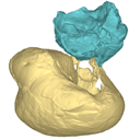
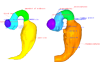
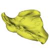
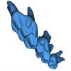
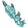
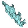
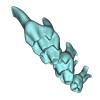
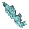
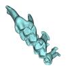
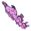
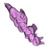
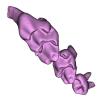
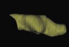
This contribution contains the 3D model described and figured in the following publication: Billet G., Germain D., Ruf I., Muizon C. de, Hautier L. 2013. The inner ear of Megatherium and the evolution of the vestibular system in sloths. Journal of Anatomy 123:557-567, DOI: 10.1111/joa.12114.
Megatherium americanum MNHN.F.PAM276 View specimen

|
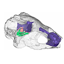
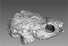
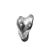
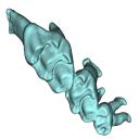
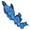
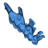
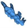
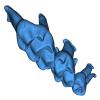
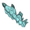
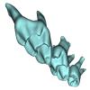
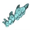
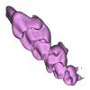
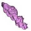
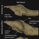
M3#14This model corresponds to a virtually reconstructed bony labyrinth of the right inner ear of the skull MNHN-F-PAM 276, attributed to the extinct giant ground sloth Megatherium americanum. The fossil comes from Pleistocene deposits at Rio Salado (Prov. Buenos Aires, Argentina). The bony labyrinth of Megatherium shows semicircular canals that are proportionally much larger than in the modern two-toed and three-toed sloths. The cochlea in Megatherium shows 2.5 turns, which is a rather high value within Xenarthra. Overall, the shape of the bony labyrinth of Megatherium resembles more that of extant armadillos than that of its extant sloth relatives. Type: "3D_surfaces"doi: 10.18563/m3.sf14 state:published |
Download 3D surface file |
This contribution contains the 3D reconstruction of Canariomys bravoi, described and figured in the following publication: Michaux J., Hautier L., Hutterer R., Lebrun R., Guy F., García-Talavera F., 2012 : Body shape and life style of the extinct rodent Canariomys bravoi (Mammalia, Murinae) from Tenerife, Canary Islands (Spain). Comptes Rendus Palevol 11 (7), 485-494. DOI: 10.1016/j.crpv.2012.06.004
Canariomys bravoi TFMCV872-873 View specimen

|
M3#6This file contains the 3D reconstruction of Canariomys bravoi, described and figured in the following publication: Michaux J., Hautier L., Hutterer R., Lebrun R., Guy F., García-Talavera F., 2012 : Body shape and life style of the extinct rodent Canariomys bravoi (Mammalia, Murinae) from Tenerife, Canary Islands (Spain). Comptes Rendus Palevol 11 (7), 485-494. Type: "3D_surfaces"doi: 10.18563/m3.sf6 state:published |
Download 3D surface file |
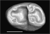
This contribution contains the 3D models described and figured in the following publication: Tabuce R., Marandat B., Adnet S., Gernelle K., Girard F., Marivaux L., Solé F., Schnyder J., Steurbaut E., Storme J.-Y., Vianey-Liaud M., Yans J. (2025). European mammal turnover driven by a global rapid warming event preceding the Paleocene-Eocene Thermal Maximum. PNAS. https://doi.org/10.1073/pnas.2505795122
Acritoparamys aff. atavus UM-ALB-41 View specimen
|
M3#17653D digital model Type: "3D_surfaces"doi: 10.18563/m3.sf.1765 state:published |
Download 3D surface file |
Acritoparamys aff. atavus UM-ALB-42 View specimen
|
M3#1766m1 (right) Type: "3D_surfaces"doi: 10.18563/m3.sf.1766 state:published |
Download 3D surface file |
Acritoparamys aff. atavus UM-ALB-43 View specimen
|
M3#1767M3 (right) Type: "3D_surfaces"doi: 10.18563/m3.sf.1767 state:published |
Download 3D surface file |
indet. indet. UM-ALB-7 View specimen
|
M3#1768M1or2 (left) Type: "3D_surfaces"doi: 10.18563/m3.sf.1768 state:published |
Download 3D surface file |
Arcius cf. rougieri UM-ALB-3 View specimen
|
M3#1769m2 (left) Type: "3D_surfaces"doi: 10.18563/m3.sf.1769 state:published |
Download 3D surface file |
Arfia sp. UM-ALB-2 View specimen
|
M3#1770M1or2 (right) Type: "3D_surfaces"doi: 10.18563/m3.sf.1770 state:published |
Download 3D surface file |
Bustylus sp. UM-ALB-37 View specimen
|
M3#1771M1 (left) Type: "3D_surfaces"doi: 10.18563/m3.sf.1771 state:published |
Download 3D surface file |
?Corbarimys sp. UM-ALB-44 View specimen
|
M3#1772M1or2 (left) Type: "3D_surfaces"doi: 10.18563/m3.sf.1772 state:published |
Download 3D surface file |
indet. indet. UM-ALB-26 View specimen
|
M3#1773upper molar (right) Type: "3D_surfaces"doi: 10.18563/m3.sf.1773 state:published |
Download 3D surface file |
indet. indet. UM-ALB-39 View specimen
|
M3#1774m1or2 (left) Type: "3D_surfaces"doi: 10.18563/m3.sf.1774 state:published |
Download 3D surface file |
Paschatherium marianae UM-ALB-4 View specimen
|
M3#1775P4 (right) Type: "3D_surfaces"doi: 10.18563/m3.sf.1775 state:published |
Download 3D surface file |
Paschatherium marianae UM-ALB-5 View specimen
|
M3#1776DP4 (right) Type: "3D_surfaces"doi: 10.18563/m3.sf.1776 state:published |
Download 3D surface file |
Paschatherium marianae UM-ALB-8 View specimen
|
M3#1777mandible with m2 and talonid of m1 (left) Type: "3D_surfaces"doi: 10.18563/m3.sf.1777 state:published |
Download 3D surface file |
Paschatherium marianae UM-ALB-10 View specimen
|
M3#1778M3 (righ Type: "3D_surfaces"doi: 10.18563/m3.sf.1778 state:published |
Download 3D surface file |
Paschatherium marianae UM-ALB-22 View specimen

|
M3#1779m3 (right) Type: "3D_surfaces"doi: 10.18563/m3.sf.1779 state:published |
Download 3D surface file |
Paschatherium marianae UM-ALB-33 View specimen
|
M3#1780M2 (right) Type: "3D_surfaces"doi: 10.18563/m3.sf.1780 state:published |
Download 3D surface file |
Peratherium sp. UM-ALB-12 View specimen
|
M3#1781?m2 (left) Type: "3D_surfaces"doi: 10.18563/m3.sf.1781 state:published |
Download 3D surface file |
Peratherium sp. UM-ALB-23 View specimen
|
M3#1782?M2 (right) Type: "3D_surfaces"doi: 10.18563/m3.sf.1782 state:published |
Download 3D surface file |
Peratherium sp. UM-ALB-25 View specimen
|
M3#1783?M3 (left) Type: "3D_surfaces"doi: 10.18563/m3.sf.1783 state:published |
Download 3D surface file |
Plagioctenodon cf. dormaalensis UM-ALB-16 View specimen
|
M3#1784M1or2 (right) Type: "3D_surfaces"doi: 10.18563/m3.sf.1784 state:published |
Download 3D surface file |
Plagioctenodon cf. dormaalensis UM-ALB-18 View specimen
|
M3#1785P4 (right) Type: "3D_surfaces"doi: 10.18563/m3.sf.1785 state:published |
Download 3D surface file |
gen. nov. sp. nov. UM-ALB-27 View specimen
|
M3#1786M1or2 (left) Type: "3D_surfaces"doi: 10.18563/m3.sf.1786 state:published |
Download 3D surface file |
Teilhardimys cf. reisi UM-ALB-36a View specimen
|
M3#1787M2 (right) Type: "3D_surfaces"doi: 10.18563/m3.sf.1787 state:published |
Download 3D surface file |
Teilhardimys cf. reisi UM-ALB-36b View specimen
|
M3#1788M1 (right) Type: "3D_surfaces"doi: 10.18563/m3.sf.1788 state:published |
Download 3D surface file |
Wyonycteris sp. UM-ALB-19 View specimen
|
M3#1789M1or2 (right) Type: "3D_surfaces"doi: 10.18563/m3.sf.1789 state:published |
Download 3D surface file |
This contribution contains the 3D models described and figured in the following publication: Georgalis, G.L., K.T. Smith, L. Marivaux, A. Herrel, E.M. Essid, H.K. Ammar, W. Marzougui, R. Temani and R. Tabuce. 2024. The world’s largest worm lizard: a new giant trogonophid (Squamata: Amphisbaenia) with extreme dental adaptations from the Eocene of Chambi, Tunisia. Zoological Journal of the Linnean Society. https://doi.org/10.1093/zoolinnean/zlae133
Terastiodontosaurus marcelosanchezi ONM CBI-1-645 View specimen
|
M3#1561Holotype maxilla ONM CBI-1-645 of Terastiodontosaurus marcelosanchezi from the Eocene of Chambi Type: "3D_surfaces"doi: 10.18563/m3.sf.1561 state:published |
Download 3D surface file |
Terastiodontosaurus marcelosanchezi ONM CBI-1-646 View specimen
|
M3#1560Paratype dentary ONM CBI-1-646 of Terastiodontosaurus marcelosanchezi from the Eocene of Chambi Type: "3D_surfaces"doi: 10.18563/m3.sf.1560 state:published |
Download 3D surface file |
Terastiodontosaurus marcelosanchezi ONM CBI-1-648 View specimen
|
M3#1562Maxilla ONM CBI-1-648 of Terastiodontosaurus marcelosanchezi from the Eocene of Chambi Type: "3D_surfaces"doi: 10.18563/m3.sf.1562 state:published |
Download 3D surface file |
Terastiodontosaurus marcelosanchezi ONM CBI-1-649 View specimen
|
M3#1559Maxilla ONM CBI-1-649 of Terastiodontosaurus marcelosanchezi from the Eocene of Chambi Type: "3D_surfaces"doi: 10.18563/m3.sf.1559 state:published |
Download 3D surface file |
Terastiodontosaurus marcelosanchezi ONM CBI-1-650 View specimen
|
M3#1563Maxilla ONM CBI-1-650 of Terastiodontosaurus marcelosanchezi from the Eocene of Chambi Type: "3D_surfaces"doi: 10.18563/m3.sf.1563 state:published |
Download 3D surface file |
Terastiodontosaurus marcelosanchezi ONM CBI-1-651 View specimen
|
M3#1564Maxilla ONM CBI-1-651 of Terastiodontosaurus marcelosanchezi from the Eocene of Chambi Type: "3D_surfaces"doi: 10.18563/m3.sf.1564 state:published |
Download 3D surface file |
Terastiodontosaurus marcelosanchezi ONM CBI-1-653 View specimen
|
M3#1565Maxilla ONM CBI-1-653 of Terastiodontosaurus marcelosanchezi from the Eocene of Chambi Type: "3D_surfaces"doi: 10.18563/m3.sf.1565 state:published |
Download 3D surface file |
Terastiodontosaurus marcelosanchezi ONM CBI-1-654 View specimen
|
M3#1576Maxilla ONM CBI-1-654 of Terastiodontosaurus marcelosanchezi from the Eocene of Chambi Type: "3D_surfaces"doi: 10.18563/m3.sf.1576 state:published |
Download 3D surface file |
Terastiodontosaurus marcelosanchezi ONM CBI-1-657 View specimen
|
M3#1566Dentary ONM CBI-1-657 of Terastiodontosaurus marcelosanchezi from the Eocene of Chambi Type: "3D_surfaces"doi: 10.18563/m3.sf.1566 state:published |
Download 3D surface file |
Terastiodontosaurus marcelosanchezi ONM CBI-1-658 View specimen
|
M3#1567Premaxilla ONM CBI-1-658 of Terastiodontosaurus marcelosanchezi from the Eocene of Chambi Type: "3D_surfaces"doi: 10.18563/m3.sf.1567 state:published |
Download 3D surface file |
Terastiodontosaurus marcelosanchezi ONM CBI-1-659 View specimen
|
M3#1568Dentary ONM CBI-1-659 of Terastiodontosaurus marcelosanchezi from the Eocene of Chambi Type: "3D_surfaces"doi: 10.18563/m3.sf.1568 state:published |
Download 3D surface file |
Terastiodontosaurus marcelosanchezi ONM CBI-1-660 View specimen
|
M3#1569Dentary ONM CBI-1-660 of Terastiodontosaurus marcelosanchezi from the Eocene of Chambi Type: "3D_surfaces"doi: 10.18563/m3.sf.1569 state:published |
Download 3D surface file |
Terastiodontosaurus marcelosanchezi ONM CBI-1-661 View specimen
|
M3#1570Dentary ONM CBI-1-661 of Terastiodontosaurus marcelosanchezi from the Eocene of Chambi Type: "3D_surfaces"doi: 10.18563/m3.sf.1570 state:published |
Download 3D surface file |
Terastiodontosaurus marcelosanchezi ONM CBI-1-668 View specimen
|
M3#1571Dentary ONM CBI-1-668 of Terastiodontosaurus marcelosanchezi from the Eocene of Chambi Type: "3D_surfaces"doi: 10.18563/m3.sf.1571 state:published |
Download 3D surface file |
Terastiodontosaurus marcelosanchezi ONM CBI-1-670 View specimen
|
M3#1572Dentary ONM CBI-1-670 of Terastiodontosaurus marcelosanchezi from the Eocene of Chambi Type: "3D_surfaces"doi: 10.18563/m3.sf.1572 state:published |
Download 3D surface file |
Terastiodontosaurus marcelosanchezi ONM CBI-1-672 View specimen
|
M3#1573Premaxilla ONM CBI-1-672 of Terastiodontosaurus marcelosanchezi from the Eocene of Chambi Type: "3D_surfaces"doi: 10.18563/m3.sf.1573 state:published |
Download 3D surface file |
Terastiodontosaurus marcelosanchezi ONM CBI-1-711 View specimen
|
M3#1574Premaxilla ONM CBI-1-711 of Terastiodontosaurus marcelosanchezi from the Eocene of Chambi Type: "3D_surfaces"doi: 10.18563/m3.sf.1574 state:published |
Download 3D surface file |
Todrasaurus gheerbranti UM THR 407 View specimen
|
M3#1575Holotype dentary UM THR 407 of Todrasaurus gheerbranti Type: "3D_surfaces"doi: 10.18563/m3.sf.1575 state:published |
Download 3D surface file |
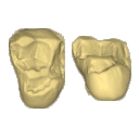
This contribution contains 3D models of upper molar rows of house mice (Mus musculus domesticus) belonging to Western European commensal and Sub-Antarctic feral populations. These two groups are characterized by different patterns of wear and alignment of the three molars along the row, related to contrasted masticatory demand in relation with their diet. These models are analyzed in the following publication: Renaud et al 2023, “Molar wear in house mice, insight into diet preferences at an ecological time scale?”, https://doi.org/10.1093/biolinnean/blad091
Mus musculus G09_06 View specimen
|
M3#1166right upper molar row Type: "3D_surfaces"doi: 10.18563/m3.sf.1166 state:published |
Download 3D surface file |
Mus musculus G09_10 View specimen
|
M3#1168right upper molar row Type: "3D_surfaces"doi: 10.18563/m3.sf.1168 state:published |
Download 3D surface file |
Mus musculus G09_15 View specimen
|
M3#1169right upper molar row Type: "3D_surfaces"doi: 10.18563/m3.sf.1169 state:published |
Download 3D surface file |
Mus musculus G09_16 View specimen
|
M3#1170right upper molar row Type: "3D_surfaces"doi: 10.18563/m3.sf.1170 state:published |
Download 3D surface file |
Mus musculus G09_17 View specimen
|
M3#1171right upper molar row Type: "3D_surfaces"doi: 10.18563/m3.sf.1171 state:published |
Download 3D surface file |
Mus musculus G09_21 View specimen
|
M3#1172right upper molar row Type: "3D_surfaces"doi: 10.18563/m3.sf.1172 state:published |
Download 3D surface file |
Mus musculus G09_26 View specimen
|
M3#1173right upper molar row Type: "3D_surfaces"doi: 10.18563/m3.sf.1173 state:published |
Download 3D surface file |
Mus musculus G09_27 View specimen
|
M3#1174right upper molar row Type: "3D_surfaces"doi: 10.18563/m3.sf.1174 state:published |
Download 3D surface file |
Mus musculus G09_29 View specimen
|
M3#1175right upper molar row Type: "3D_surfaces"doi: 10.18563/m3.sf.1175 state:published |
Download 3D surface file |
Mus musculus G09_65 View specimen
|
M3#1176right upper molar row Type: "3D_surfaces"doi: 10.18563/m3.sf.1176 state:published |
Download 3D surface file |
Mus musculus G09_66 View specimen
|
M3#1177right upper molar row Type: "3D_surfaces"doi: 10.18563/m3.sf.1177 state:published |
Download 3D surface file |
Mus musculus G93_03 View specimen
|
M3#1178right upper molar row Type: "3D_surfaces"doi: 10.18563/m3.sf.1178 state:published |
Download 3D surface file |
Mus musculus G93_04 View specimen
|
M3#1179right upper molar row Type: "3D_surfaces"doi: 10.18563/m3.sf.1179 state:published |
Download 3D surface file |
Mus musculus G93_10 View specimen
|
M3#1180right upper molar row Type: "3D_surfaces"doi: 10.18563/m3.sf.1180 state:published |
Download 3D surface file |
Mus musculus G93_11 View specimen
|
M3#1181right upper molar row Type: "3D_surfaces"doi: 10.18563/m3.sf.1181 state:published |
Download 3D surface file |
Mus musculus G93_13 View specimen
|
M3#1182right upper molar row Type: "3D_surfaces"doi: 10.18563/m3.sf.1182 state:published |
Download 3D surface file |
Mus musculus G93_14 View specimen
|
M3#1183right upper molar row Type: "3D_surfaces"doi: 10.18563/m3.sf.1183 state:published |
Download 3D surface file |
Mus musculus G93_15 View specimen
|
M3#1184right upper molar row Type: "3D_surfaces"doi: 10.18563/m3.sf.1184 state:published |
Download 3D surface file |
Mus musculus G93_24 View specimen
|
M3#1185left molar row Type: "3D_surfaces"doi: 10.18563/m3.sf.1185 state:published |
Download 3D surface file |
Mus musculus Tourch_7819 View specimen
|
M3#1186right upper molar row Type: "3D_surfaces"doi: 10.18563/m3.sf.1186 state:published |
Download 3D surface file |
Mus musculus G93_25 View specimen
|
M3#1187right upper molar row Type: "3D_surfaces"doi: 10.18563/m3.sf.1187 state:published |
Download 3D surface file |
Mus musculus Tourch_7821 View specimen
|
M3#1188right upper molar row Type: "3D_surfaces"doi: 10.18563/m3.sf.1188 state:published |
Download 3D surface file |
Mus musculus Tourch_7839 View specimen
|
M3#1189right upper molar row Type: "3D_surfaces"doi: 10.18563/m3.sf.1189 state:published |
Download 3D surface file |
Mus musculus Tourch_7873 View specimen
|
M3#1190right upper molar row Type: "3D_surfaces"doi: 10.18563/m3.sf.1190 state:published |
Download 3D surface file |
Mus musculus Tourch_7877 View specimen
|
M3#1196right upper molar row Type: "3D_surfaces"doi: 10.18563/m3.sf.1196 state:published |
Download 3D surface file |
Mus musculus Tourch_7922 View specimen
|
M3#1191right upper molar row Type: "3D_surfaces"doi: 10.18563/m3.sf.1191 state:published |
Download 3D surface file |
Mus musculus Tourch_7923 View specimen
|
M3#1192right upper molar row Type: "3D_surfaces"doi: 10.18563/m3.sf.1192 state:published |
Download 3D surface file |
Mus musculus Tourch_7925 View specimen
|
M3#1193right upper molar row Type: "3D_surfaces"doi: 10.18563/m3.sf.1193 state:published |
Download 3D surface file |
Mus musculus Tourch_7927 View specimen
|
M3#1194right upper molar row Type: "3D_surfaces"doi: 10.18563/m3.sf.1194 state:published |
Download 3D surface file |
Mus musculus Tourch_7932 View specimen
|
M3#1195right upper molar row Type: "3D_surfaces"doi: 10.18563/m3.sf.1195 state:published |
Download 3D surface file |
The present 3D Dataset contains the 3D model analyzed in Gaetano, L. C., Abdala, F., Seoane, F. D., Tartaglione, A., Schulz, M., Otero, A., Leardi, J. M., Apaldetti, C., Krapovickas, V., and Steinbach, E. 2021. A new cynodont from the Upper Triassic Los Colorados Formation (Argentina, South America) reveals a novel paleobiogeographic context for mammalian ancestors. Scientific Reports.
Tessellatia bonapartei PULR-V121 View specimen
|
M3#9603D surface model of PULR-V121 Type: "3D_surfaces"doi: 10.18563/m3.sf.960 state:published |
Download 3D surface file |
The present 3D Dataset contains 26 3D models analyzed in the study: On the “cartilaginous rider” in the endocasts of turtle brain cavities, published by the authors in the journal Vertebrate Zoology.
Annemys sp. IVPP-V-18106 View specimen

|
M3#7723D surface(s) file to specimen IVPP-V-18106 Type: "3D_surfaces"doi: 10.18563/m3.sf.772 state:published |
Download 3D surface file |
Apalone spinifera FMNH 22178 View specimen

|
M3#7733D surface(s) file to specimen FMNH 22178 Type: "3D_surfaces"doi: 10.18563/m3.sf.773 state:published |
Download 3D surface file |
Caretta caretta NHMUK1940.3.15.1 View specimen
|
M3#7863D surface(s) file to specimen NHMUK1940.3.15.1 Type: "3D_surfaces"doi: 10.18563/m3.sf.786 state:published |
Download 3D surface file |
Chelodina reimanni ZMB 49659 View specimen

|
M3#7743D surface(s) file to specimen ZMB 49659 Type: "3D_surfaces"doi: 10.18563/m3.sf.774 state:published |
Download 3D surface file |
Chelonia mydas ZMB-37416MS View specimen
|
M3#7753D surface(s) file to specimen ZMB-37416MS Type: "3D_surfaces"doi: 10.18563/m3.sf.775 state:published |
Download 3D surface file |
Cuora amboinensis NHMUK69.42.145_4 View specimen
|
M3#7763D surface(s) file to specimen NHMUK69.42.145_4 Type: "3D_surfaces"doi: 10.18563/m3.sf.776 state:published |
Download 3D surface file |
Emydura subglobosa IW92 View specimen
|
M3#7773D surface(s) file to specimen IW92 Type: "3D_surfaces"doi: 10.18563/m3.sf.777 state:published |
Download 3D surface file |
Eubaena cephalica DMNH 96004 View specimen
|
M3#7783D surface(s) file to specimen DMNH 96004 Type: "3D_surfaces"doi: 10.18563/m3.sf.778 state:published |
Download 3D surface file |
Gopherus berlandieri AMNH-73816 View specimen
|
M3#7793D surface(s) file to specimen AMNH-73816 Type: "3D_surfaces"doi: 10.18563/m3.sf.779 state:published |
Download 3D surface file |
Kinixys belliana AMNH-10028 View specimen
|
M3#7803D surface(s) file to specimen AMNH-10028 Type: "3D_surfaces"doi: 10.18563/m3.sf.780 state:published |
Download 3D surface file |
Macrochelys temminckii GPIT-PV-79430 View specimen

|
M3#7813D surface(s) file to specimen GPIT-PV-79430 Type: "3D_surfaces"doi: 10.18563/m3.sf.781 state:published |
Download 3D surface file |
Malacochersus tornieri SMF-58702 View specimen
|
M3#7873D surface(s) file to specimen SMF-58702 Type: "3D_surfaces"doi: 10.18563/m3.sf.787 state:published |
Download 3D surface file |
Naomichelys speciosa FMNH-PR-273 View specimen
|
M3#7823D surface(s) file to specimen FMNH-PR-273 Type: "3D_surfaces"doi: 10.18563/m3.sf.782 state:published |
Download 3D surface file |
Pelodiscus sinensis IW576-2 View specimen
|
M3#7833D surface(s) file to specimen IW576-2 Type: "3D_surfaces"doi: 10.18563/m3.sf.783 state:published |
Download 3D surface file |
Platysternon megacephalum SMF-69684 View specimen
|
M3#7843D surface(s) file to specimen SMF-69684 Type: "3D_surfaces"doi: 10.18563/m3.sf.784 state:published |
Download 3D surface file |
Podocnemis unifilis SMF-55470 View specimen
|
M3#7853D surface(s) file to specimen SMF-55470 Type: "3D_surfaces"doi: 10.18563/m3.sf.785 state:published |
Download 3D surface file |
Proganochelys quenstedtii MB 1910.45.2 View specimen
|
M3#7883D surface(s) file to specimen MB 1910.45.2 Type: "3D_surfaces"doi: 10.18563/m3.sf.788 state:published |
Download 3D surface file |
Proganochelys quenstedtii SMNS 16980 View specimen
|
M3#7893D surface(s) file to specimen SMNS 16980 Type: "3D_surfaces"doi: 10.18563/m3.sf.789 state:published |
Download 3D surface file |
Rhinochelys pulchriceps CAMSM_B55775 View specimen
|
M3#7903D surface(s) file to specimen CAMSM_B55775 Type: "3D_surfaces"doi: 10.18563/m3.sf.790 state:published |
Download 3D surface file |
Rhinoclemmys funereal YPM12174 View specimen
|
M3#7913D surface(s) file to specimen YPM12174 Type: "3D_surfaces"doi: 10.18563/m3.sf.791 state:published |
Download 3D surface file |
Sandownia harrisi MIWG3480 View specimen
|
M3#7923D surface(s) file to specimen MIWG3480 Type: "3D_surfaces"doi: 10.18563/m3.sf.792 state:published |
Download 3D surface file |
Testudo graeca YPM14342 View specimen
|
M3#7933D surface(s) file to specimen YPM14342 Type: "3D_surfaces"doi: 10.18563/m3.sf.793 state:published |
Download 3D surface file |
Testudo hermanni AMNH134518 View specimen
|
M3#7943D surface(s) file to specimen AMNH134518 Type: "3D_surfaces"doi: 10.18563/m3.sf.794 state:published |
Download 3D surface file |
Trachemys scripta NN View specimen
|
M3#7953D surface(s) file to specimen Trachemys scripta Type: "3D_surfaces"doi: 10.18563/m3.sf.795 state:published |
Download 3D surface file |
Xinjiangchelys radiplicatoides IVPP V9539 View specimen
|
M3#7963D surface(s) file to specimen IVPP V9539 Type: "3D_surfaces"doi: 10.18563/m3.sf.796 state:published |
Download 3D surface file |
Chelydra serpentina UFR VP1 View specimen

|
M3#801Brain endocast Type: "3D_surfaces"doi: 10.18563/m3.sf.801 state:published |
Download 3D surface file |
This contribution contains the 3D models of the fossil teeth of a small-bodied platyrrhine primate, Neosaimiri cf. fieldsi (Cebinae, Cebidae, Platyrrhini) discovered from Laventan deposits (late Middle Miocene) of Peruvian Amazonia, San Martín Department (TAR-31: Tarapoto/Juan Guerra vertebrate fossil-bearing locus n°31). These fossils were described and figured in the following publication: Marivaux et al. (2020), New record of Neosaimiri (Cebidae, Platyrrhini) from the late Middle Miocene of Peruvian Amazonia. Journal of Human Evolution. https://doi.org/10.1016/j.jhevol.2020.102835
Neosaimiri cf. fieldsi MUSM-3888 View specimen
|
M3#538MUSM-3888, right m3 of Neosaimiri cf. fieldsi. Type: "3D_surfaces"doi: 10.18563/m3.sf.538 state:published |
Download 3D surface file |
Neosaimiri cf. fieldsi MUSM-3890 View specimen
|
M3#540MUSM-3890, left dp2 of Neosaimiri cf. fieldsi. Type: "3D_surfaces"doi: 10.18563/m3.sf.540 state:published |
Download 3D surface file |
Neosaimiri cf. fieldsi MUSM-3895 View specimen
|
M3#541MUSM-3895, right DC1 of Neosaimiri cf. fieldsi. Type: "3D_surfaces"doi: 10.18563/m3.sf.541 state:published |
Download 3D surface file |
Neosaimiri cf. fieldsi MUSM-3891 View specimen
|
M3#542MUSM-3891, lingual part of a fragmentary right M1 or M2 of Neosaimiri cf. fieldsi. Type: "3D_surfaces"doi: 10.18563/m3.sf.542 state:published |
Download 3D surface file |
Neosaimiri cf. fieldsi MUSM-3892 View specimen
|
M3#543MUSM-3892, distobuccal part of a fragmentary right upper molar (metacone region) of Neosaimiri cf. fieldsi. Type: "3D_surfaces"doi: 10.18563/m3.sf.543 state:published |
Download 3D surface file |
Neosaimiri cf. fieldsi MUSM-3893 View specimen
|
M3#544MUSM-3893, buccal part of a fragmentary right P3 or P4 of Neosaimiri cf. fieldsi. Type: "3D_surfaces"doi: 10.18563/m3.sf.544 state:published |
Download 3D surface file |
Neosaimiri cf. fieldsi MUSM-3894 View specimen
|
M3#545MUSM-3894, lingual part of a fragmentary left P3 or P4 of Neosaimiri cf. fieldsi. Type: "3D_surfaces"doi: 10.18563/m3.sf.545 state:published |
Download 3D surface file |
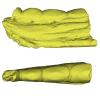
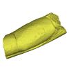
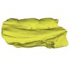
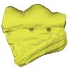
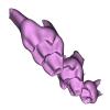
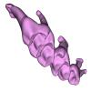
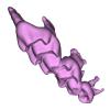
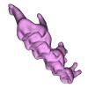
The present Dataset contains the 3D model of the male genital organs of greater horseshoe bat, Rhinolophus ferrumequinum. This is the first detailed 3D structure of the soft-tissue genital organs of bats. The 3D model was generated using microCT and techniques of virtual reconstruction.
Rhinolophus ferrumequinum JP18-006 View specimen
|
M3#521The genital organs of male greater horseshoe bat. Type: "3D_surfaces"doi: 10.18563/m3.sf.521 state:published |
Download 3D surface file |
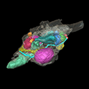
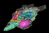
The present 3D Dataset contains the 3D models analyzed in the following publication: Paulina-Carabajal, A., Ezcurra, M., Novas, F., 2019. New information on the braincase and endocranial morphology of the Late Triassic neotheropod Zupaysaurus rougieri using Computed Tomography data. Journal of Vertebrate Paleontology. https://doi.org/10.1080/02724634.2019.1630421
Zupaysaurus rougieri PULR 076 View specimen
|
M3#424The Zip contains 3 files, which correspond to: PULR_076-M1: Zupaysaurus rougieri skull, braincase and cranial endocast PULR_076-M2: Zupaysaurus rougieri braincase PULR_076-M1: Zupaysaurus rougieri brain and inner ear Type: "3D_surfaces"doi: 10.18563/m3.sf.424 state:published |
Download 3D surface file |
This contribution contains the 3D models described and figured in the following publication: Tissier et al. (in prep.).
Sellamynodon zimborensis UBB MPS 15795 View specimen
|
M3#297Incomplete skull with left M3. Type: "3D_surfaces"doi: 10.18563/m3.sf.297 state:published |
Download 3D surface file |
Sellamynodon zimborensis UBB MPS 15795 View specimen
|
M3#298Mandible with complete molar and premolar rows, lacking symphysis. Type: "3D_surfaces"doi: 10.18563/m3.sf.298 state:published |
Download 3D surface file |
Amynodontopsis aff. bodei UBB MPS V545 View specimen
|
M3#299Maxillary fragment with M1-3. Type: "3D_surfaces"doi: 10.18563/m3.sf.299 state:published |
Download 3D surface file |
Amynodontopsis aff. bodei UBB MPS V546 View specimen
|
M3#300Unworn m1/2 on mandible fragment. Type: "3D_surfaces"doi: 10.18563/m3.sf.300 state:published |
Download 3D surface file |
This contribution contains the 3D models of the isolated teeth of Canaanimico amazonensis, a new stem platyrrhine primate, described and figured in the following publication: Marivaux et al. (2016), Neotropics provide insights into the emergence of New World monkeys: new dental evidence from the late Oligocene of Peruvian Amazonia. Journal of Human Evolution. http://dx.doi.org/10.1016/j.jhevol.2016.05.011
Canaanimico amazonensis MUSM-2499 View specimen
|
M3#2893D model of left upper M2 Type: "3D_surfaces"doi: 10.18563/m3.sf.289 state:published |
Download 3D surface file |
Canaanimico amazonensis MUSM-2500 View specimen
|
M3#2903D model of left upper M1 (lingual part) Type: "3D_surfaces"doi: 10.18563/m3.sf.290 state:published |
Download 3D surface file |
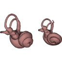
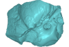
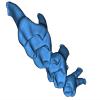
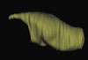
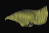
The present 3D Dataset contains the 3D models analyzed in Velazco P. M., Grohé C. 2017. Comparative anatomy of the bony labyrinth of the bats Platalina genovensium (Phyllostomidae, Lonchophyllinae) and Tomopeas ravus (Molossidae, Tomopeatinae). Biotempo 14(2).
Platalina genovensium 278520 View specimen

|
M3#276Right bony labyrinth surface positioned (.PLY) Labels associated (.FLG) Type: "3D_surfaces"doi: 10.18563/m3.sf.276 state:published |
Download 3D surface file |
Tomopeas ravus 278525 View specimen

|
M3#277Right bony labyrinth surface (.PLY) Labels associated (.FLG) Type: "3D_surfaces"doi: 10.18563/m3.sf.277 state:published |
Download 3D surface file |
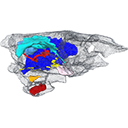
The present 3D Dataset contains the 3D models analyzed in: "a giant dapediid from the Late Triassic of Switzerland and insights into neopterygian phylogeny", Royal Society Open Science, https://doi.org/10.1098/rsos.180497
Scopulipiscis saxciput PIMUZ A/I 3026 View specimen

|
M3#1773D surfaces of the skull and endocranial spaces inside neurocranium, including the aortic canal, braincase, fossa bridgei, lateral cranial canal, nerves and other passageways, notochord, posterior myodome, and right semicircular canals. Type: "3D_surfaces"doi: 10.18563/m3.sf.177 state:published |
Download 3D surface file |

|
M3#178Scan of the neurocranium of PIMUZ A/I 3026 Type: "3D_CT"doi: 10.18563/m3.sf.178 state:published |
Download CT data |


