Explodable 3D Dog Skull for Veterinary Education
3D models of Ocnotherium skull
3D models of Kalakocetus, the earliest Cetacea
3D GM dataset of bird skeletal variation
Skeletal embryonic development in the catshark
Bony connexions of the petrosal bone of extant hippos
bony labyrinth (14) , inner ear (11) , geometric morphometrics (10) , CT-scan (10) , Eocene (10) , Micro-CT (9) , Miocene (8)
Lionel Hautier (24) , Maëva Judith Orliac (23) , Laurent Marivaux (18) , Renaud Lebrun (14) , Rodolphe Tabuce (14) , Pierre-Olivier Antoine (13) , Bastien Mennecart (13)
|
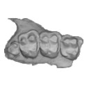
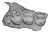
3D models related to the publication: Sniffing out morphological convergence in the turbinal complex of myrmecophagous placentals.Mark Wright
Published online: 21/11/2024 |
|
M3#1536Turbinals of Priodontes maximus Type: "3D_surfaces"doi: 10.18563/m3.sf.1536 state:published |
Download 3D surface file |
Dasypus pilosus NHMUK 94-10-1-13 View specimen
|
M3#1537Turbinals of Dasypus pilosus Type: "3D_surfaces"doi: 10.18563/m3.sf.1537 state:published |
Download 3D surface file |
Dasypus novemcinctus AMNH 263287 View specimen
|
M3#1538Turbinals of Dasypus novemcinctus Type: "3D_surfaces"doi: 10.18563/m3.sf.1538 state:published |
Download 3D surface file |
Bradypus tridactylus UM 789N View specimen
|
M3#1539Turbinals of Bradypus tridactylus Type: "3D_surfaces"doi: 10.18563/m3.sf.1539 state:published |
Download 3D surface file |
Choloepus didactylus UM 767V View specimen
|
M3#1540Turbinals of Choloepus didactylus Type: "3D_surfaces"doi: 10.18563/m3.sf.1540 state:published |
Download 3D surface file |
Cyclopes didactylus NHMUK 88-8-8-14 View specimen
|
M3#1541Turbinals of Cyclopes didactylus Type: "3D_surfaces"doi: 10.18563/m3.sf.1541 state:published |
Download 3D surface file |
Myrmecophaga tridactyla UM 065V View specimen
|
M3#1542Turbinals of Myrmecophaga tridactyla Type: "3D_surfaces"doi: 10.18563/m3.sf.1542 state:published |
Download 3D surface file |
Tamandua tetradactyla NHMUK 3-7-7-135 View specimen
|
M3#1543Turbinals of Tamandua tetradactyla Type: "3D_surfaces"doi: 10.18563/m3.sf.1543 state:published |
Download 3D surface file |
Tamandua mexicana NHMUK 79-1-6-1 View specimen
|
M3#1544Turbinals of Tamandua mexicana Type: "3D_surfaces"doi: 10.18563/m3.sf.1544 state:published |
Download 3D surface file |
Orycteropus afer NHMUK 2-9-9-58 View specimen
|
M3#1545Turbinals of Orycteropus afer Type: "3D_surfaces"doi: 10.18563/m3.sf.1545 state:published |
Download 3D surface file |
Tenrec eucaudatus UM N439 View specimen
|
M3#1546Turbinals of Tenrec eucaudatus Type: "3D_surfaces"doi: 10.18563/m3.sf.1546 state:published |
Download 3D surface file |
Elephantulus rozeti UM N227 View specimen
|
M3#1547Turbinals of Elephantulus rozeti Type: "3D_surfaces"doi: 10.18563/m3.sf.1547 state:published |
Download 3D surface file |
Phataginus tetradactyla NHMUK 1-11-21-35 View specimen
|
M3#1548Turbinals of Phataginus tetradactyla Type: "3D_surfaces"doi: 10.18563/m3.sf.1548 state:published |
Download 3D surface file |
Smutsia gigantea KMMA 25479 View specimen
|
M3#1549Turbinals of Smutsia gigantea Type: "3D_surfaces"doi: 10.18563/m3.sf.1549 state:published |
Download 3D surface file |
Manis culionensis MNHN ZM-MO 1884-1822 View specimen
|
M3#1550Turbinals of Manis culionensis Type: "3D_surfaces"doi: 10.18563/m3.sf.1550 state:published |
Download 3D surface file |
Vulpes vulpes UM N140 View specimen
|
M3#1551Turbinals of Vulpes vulpes Type: "3D_surfaces"doi: 10.18563/m3.sf.1551 state:published |
Download 3D surface file |
Alopex lagopus UM N110 View specimen
|
M3#1552Turbinals of Alopex lagopus Type: "3D_surfaces"doi: 10.18563/m3.sf.1552 state:published |
Download 3D surface file |
Felis silvestris UM N149 View specimen
|
M3#1553Turbinals of Felis sylvestris Type: "3D_surfaces"doi: 10.18563/m3.sf.1553 state:published |
Download 3D surface file |
Hyaena hyaena UM N109 View specimen
|
M3#1554Turbinals of Hyaena hyaena Type: "3D_surfaces"doi: 10.18563/m3.sf.1554 state:published |
Download 3D surface file |
Proteles cristata NHMUK 4-3-1-58 View specimen
|
M3#1555Turbinals of Proteles cristata Type: "3D_surfaces"doi: 10.18563/m3.sf.1555 state:published |
Download 3D surface file |
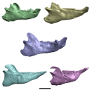
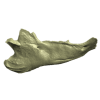
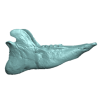
The present 3D Dataset contains the 3D models analyzed in 3D Finite Element Analysis and Geometric Morphometrics of Sloths (Xenarthra, Folivora) Mandibles Show Insights on the Dietary Specializations of Fossil Taxa. Journal of South American Earth Sciences. https://doi.org/10.1016/j.jsames.2023.104445
Mylodon darwinii CAV 379 View specimen
|
M3#1159Right hemimandible Type: "3D_surfaces"doi: 10.18563/m3.sf.1159 state:published |
Download 3D surface file |
Scelidotherium leptocephalum MNHN-M 137,722 View specimen
|
M3#1160Mandible Type: "3D_surfaces"doi: 10.18563/m3.sf.1160 state:published |
Download 3D surface file |
Glossotherium robustum MNHN-M 914 View specimen
|
M3#1161Mandible Type: "3D_surfaces"doi: 10.18563/m3.sf.1161 state:published |
Download 3D surface file |
Lestodon armatus MPAC 899 View specimen
|
M3#1162Mandible Type: "3D_surfaces"doi: 10.18563/m3.sf.1162 state:published |
Download 3D surface file |
Valgipes bucklandi NHMD.Z.M.K. 1/1845:3540 View specimen
|
M3#1163Mandible Type: "3D_surfaces"doi: 10.18563/m3.sf.1163 state:published |
Download 3D surface file |
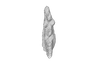
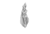
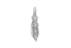
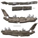
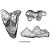
This contribution contains the 3D models of a set of Famennian conodont elements belonging to the species Icriodus alternatus analyzed in the following publication: Girard et al. 2022: Deciphering the morphological variation and its ontogenetic dynamics in the Late Devonian conodont Icriodus alternatus.
Icriodus alternatus UM BUS 031 View specimen
|
M3#887conodont element Type: "3D_surfaces"doi: 10.18563/m3.sf.887 state:published |
Download 3D surface file |
Icriodus alternatus UM BUS 032 View specimen
|
M3#888conodont element Type: "3D_surfaces"doi: 10.18563/m3.sf.888 state:published |
Download 3D surface file |
Icriodus alternatus UM BUS 033 View specimen
|
M3#889conodont element Type: "3D_surfaces"doi: 10.18563/m3.sf.889 state:published |
Download 3D surface file |
Icriodus alternatus UM BUS 034 View specimen
|
M3#890conodont element Type: "3D_surfaces"doi: 10.18563/m3.sf.890 state:published |
Download 3D surface file |
Icriodus alternatus UM BUS 035 View specimen
|
M3#891conodont element Type: "3D_surfaces"doi: 10.18563/m3.sf.891 state:published |
Download 3D surface file |
Icriodus alternatus UM BUS 036 View specimen
|
M3#892conodont element Type: "3D_surfaces"doi: 10.18563/m3.sf.892 state:published |
Download 3D surface file |
Icriodus alternatus UM BUS 037 View specimen
|
M3#893conodont element Type: "3D_surfaces"doi: 10.18563/m3.sf.893 state:published |
Download 3D surface file |
Icriodus alternatus UM BUS 038 View specimen
|
M3#894conodont element Type: "3D_surfaces"doi: 10.18563/m3.sf.894 state:published |
Download 3D surface file |
Icriodus alternatus UM BUS 039 View specimen
|
M3#895conodont element Type: "3D_surfaces"doi: 10.18563/m3.sf.895 state:published |
Download 3D surface file |
Icriodus alternatus UM BUS 040 View specimen
|
M3#896conodont element Type: "3D_surfaces"doi: 10.18563/m3.sf.896 state:published |
Download 3D surface file |
Icriodus alternatus UM BUS 041 View specimen
|
M3#897conodont element Type: "3D_surfaces"doi: 10.18563/m3.sf.897 state:published |
Download 3D surface file |
Icriodus alternatus UM BUS 042 View specimen
|
M3#898conodont element Type: "3D_surfaces"doi: 10.18563/m3.sf.898 state:published |
Download 3D surface file |
Icriodus alternatus UM BUS 043 View specimen
|
M3#899conodont element Type: "3D_surfaces"doi: 10.18563/m3.sf.899 state:published |
Download 3D surface file |
Icriodus alternatus UM BUS 044 View specimen
|
M3#900conodont element Type: "3D_surfaces"doi: 10.18563/m3.sf.900 state:published |
Download 3D surface file |
Icriodus alternatus UM BUS 045 View specimen
|
M3#901conodont element Type: "3D_surfaces"doi: 10.18563/m3.sf.901 state:published |
Download 3D surface file |
This contribution contains the 3D models described and figured in the following publications:
- Marini E., Lussu P., 2020. A virtual physical anthropology lab. Teaching in the time of coronavirus, in prep.;
- Lussu P., Bratzu D., Marini E., 2020. Cloud-based ultra close-range digital photogrammetry: validation of an approach for the effective virtual reconstruction of skeletal remains, in prep.
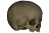
Homo sapiens MSAE 59 View specimen

|
M3#509MSAE 59 Type: "3D_surfaces"doi: 10.18563/m3.sf.509 state:published |
Download 3D surface file |
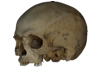
Homo sapiens MSAE 62 View specimen
|
M3#510MSAE 62 Type: "3D_surfaces"doi: 10.18563/m3.sf.510 state:published |
Download 3D surface file |
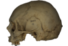
Homo sapiens MSAE 63 View specimen
|
M3#512MSAE 63 Type: "3D_surfaces"doi: 10.18563/m3.sf.512 state:published |
Download 3D surface file |
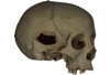
Homo sapiens MSAE 78 View specimen
|
M3#514MSAE 78 Type: "3D_surfaces"doi: 10.18563/m3.sf.514 state:published |
Download 3D surface file |
Homo sapiens MSAE 95 View specimen
|
M3#515MSAE 95 Type: "3D_surfaces"doi: 10.18563/m3.sf.515 state:published |
Download 3D surface file |
Homo sapiens MSAE 1852 View specimen
|
M3#516MSAE 1852 Type: "3D_surfaces"doi: 10.18563/m3.sf.516 state:published |
Download 3D surface file |
Homo sapiens MSAE 6426 View specimen
|
M3#517MSAE 6426 Type: "3D_surfaces"doi: 10.18563/m3.sf.517 state:published |
Download 3D surface file |
Homo sapiens MSAE 6428 View specimen
|
M3#518MSAE 6428 Type: "3D_surfaces"doi: 10.18563/m3.sf.518 state:published |
Download 3D surface file |
Homo sapiens MSAE 6992 View specimen
|
M3#519MSAE 6992 Type: "3D_surfaces"doi: 10.18563/m3.sf.519 state:published |
Download 3D surface file |
Homo sapiens MSAE 7688 View specimen
|
M3#520MSAE 7688 Type: "3D_surfaces"doi: 10.18563/m3.sf.520 state:published |
Download 3D surface file |
This contribution includes the 3D models of the reconstructed ossicular chain of the cainotheriid Caenomeryx filholi from the late Oligocene locality of Pech Desse (MP28, Quercy, France) described and figured in the publication of Assemat et al. (2020). It represents the oldest ossicular chain reconstruction for a Paleogene terrestrial artiodactyl species.
Caenomeryx filholi UM PDS 3353 View specimen
|
M3#508reconstruction of the middle ear with petrosal, bulla, stapes, incus, malleus Type: "3D_surfaces"doi: 10.18563/m3.sf.508 state:published |
Download 3D surface file |
The present 3D Dataset contains the 3D models described and figured in the following publication: Grohé C., Bonis L. de, Chaimanee Y., Chavasseau O., Rugbumrung M., Yamee C., Suraprasit K., Gibert C., Surault J., Blondel C., Jaeger J.-J. 2020. The late middle Miocene Mae Moh Basin of northern Thailand: the richest Neogene assemblage of Carnivora from Southeast Asia and a paleobiogeographic analysis of Miocene Asian carnivorans. American Museum Novitates. http://digitallibrary.amnh.org/handle/2246/7223
Siamogale bounosa MM-54 View specimen
|
M3#5053D model of the skull of Siamogale bounosa The zip file contains: - the 3D surface in PLY - the orientation files in .pos and .ori - the project in .ntw Type: "3D_surfaces"doi: 10.18563/m3.sf.505 state:published |
Download 3D surface file |
Vishnuonyx maemohensis MM-78 View specimen

|
M3#5063D model of the skull of Vishnuonyx maemohensis The zip file contains: - the 3D surface in PLY - the orientation files in .pos and .ori - the project in .ntw Type: "3D_surfaces"doi: 10.18563/m3.sf.506 state:published |
Download 3D surface file |
|
M3#5073D model of the reconstructed upper teeth of Vishnuonyx maemohensis The zip file contains: - the 3D surface in PLY - the orientation files in .pos and .ori - the project in .ntw Type: "3D_surfaces"doi: 10.18563/m3.sf.507 state:published |
Download 3D surface file |
This contribution contains the 3D model of the holotype of Chambius kasserinensis, the basalmost ‘elephant-shrew’ figured in the following publication: New remains of Chambius kasserinensis from the Eocene of Tunisia and evaluation of proposed affinities for Macroscelidea (Mammalia, Afrotheria). https://doi.org/10.1080/08912963.2017.1297433
Chambius kasserinensis CBI-1-06 View specimen
|
M3#1463D model of the holotype maxilla of Chambius kasserinensis. The 3D surface was extracted manually from the limestone matrix within AVIZO 9.2 Type: "3D_surfaces"doi: 10.18563/m3.sf.146 state:published |
Download 3D surface file |
This contribution contains 3D models of left and right house mouse (Mus musculus domesticus) inner ears analyzed in Renaud et al. (2024). The studied mice belong to four groups: wild-trapped mice, wild-derived lab offspring, a typical laboratory strain (Swiss) and hybrids between wild-derived and Swiss mice. They have been analyzed to assess the impact of mobility reduction on inner ear morphology, including patterns of divergence, levels of inter-individual variance (disparity) and intra-individual variance (fluctuating asymmetry)
Mus musculus Tourch_7819 View specimen
|
M3#1366Two bony labyrinths Type: "3D_surfaces"doi: 10.18563/m3.sf.1366 state:published |
Download 3D surface file |
Mus musculus Tourch_7821 View specimen
|
M3#1367Two bony labyrinths Type: "3D_surfaces"doi: 10.18563/m3.sf.1367 state:published |
Download 3D surface file |
Mus musculus Tourch_7839 View specimen
|
M3#1368Two bony labyrinths Type: "3D_surfaces"doi: 10.18563/m3.sf.1368 state:published |
Download 3D surface file |
Mus musculus Tourch_7873 View specimen
|
M3#1369Two bony labyrinths Type: "3D_surfaces"doi: 10.18563/m3.sf.1369 state:published |
Download 3D surface file |
Mus musculus Tourch_7877 View specimen
|
M3#1370Two bony labyrinths Type: "3D_surfaces"doi: 10.18563/m3.sf.1370 state:published |
Download 3D surface file |
Mus musculus Tourch_7922 View specimen
|
M3#1371Two bony labyrinths Type: "3D_surfaces"doi: 10.18563/m3.sf.1371 state:published |
Download 3D surface file |
Mus musculus Tourch_7923 View specimen
|
M3#1372Two bony labyrinths Type: "3D_surfaces"doi: 10.18563/m3.sf.1372 state:published |
Download 3D surface file |
Mus musculus Tourch_7925 View specimen
|
M3#1373Two bony labyrinths Type: "3D_surfaces"doi: 10.18563/m3.sf.1373 state:published |
Download 3D surface file |
Mus musculus Tourch_7927 View specimen
|
M3#1374Two bony labyrinths Type: "3D_surfaces"doi: 10.18563/m3.sf.1374 state:published |
Download 3D surface file |
Mus musculus Tourch_7932 View specimen
|
M3#1375Two bony labyrinths Type: "3D_surfaces"doi: 10.18563/m3.sf.1375 state:published |
Download 3D surface file |
Mus musculus Bal02 View specimen
|
M3#1320Two bony labyrinths Type: "3D_surfaces"doi: 10.18563/m3.sf.1320 state:published |
Download 3D surface file |
Mus musculus Bal04 View specimen
|
M3#1321Two bony labyrinths Type: "3D_surfaces"doi: 10.18563/m3.sf.1321 state:published |
Download 3D surface file |
Mus musculus Bal06 View specimen
|
M3#1322Two bony labyrinths Type: "3D_surfaces"doi: 10.18563/m3.sf.1322 state:published |
Download 3D surface file |
Mus musculus Bal08 View specimen
|
M3#1323Two bony labyrinths Type: "3D_surfaces"doi: 10.18563/m3.sf.1323 state:published |
Download 3D surface file |
Mus musculus Bal11 View specimen
|
M3#1324Two bony labyrinths Type: "3D_surfaces"doi: 10.18563/m3.sf.1324 state:published |
Download 3D surface file |
Mus musculus Bal12 View specimen
|
M3#1325Two bony labyrinths Type: "3D_surfaces"doi: 10.18563/m3.sf.1325 state:published |
Download 3D surface file |
Mus musculus Bal15 View specimen
|
M3#1326Two bony labyrinths Type: "3D_surfaces"doi: 10.18563/m3.sf.1326 state:published |
Download 3D surface file |
Mus musculus Bal16 View specimen
|
M3#1327Two bony labyrinths Type: "3D_surfaces"doi: 10.18563/m3.sf.1327 state:published |
Download 3D surface file |
Mus musculus Bal17 View specimen
|
M3#1328Two bony labyrinths Type: "3D_surfaces"doi: 10.18563/m3.sf.1328 state:published |
Download 3D surface file |
Mus musculus Bal18 View specimen
|
M3#1329Two bony labyrinths Type: "3D_surfaces"doi: 10.18563/m3.sf.1329 state:published |
Download 3D surface file |
Mus musculus Bal19 View specimen
|
M3#1330Two bony labyrinths Type: "3D_surfaces"doi: 10.18563/m3.sf.1330 state:published |
Download 3D surface file |
Mus musculus Bal20 View specimen
|
M3#1331Two bony labyrinths Type: "3D_surfaces"doi: 10.18563/m3.sf.1331 state:published |
Download 3D surface file |
Mus musculus Bal21 View specimen
|
M3#1332Two bony labyrinths Type: "3D_surfaces"doi: 10.18563/m3.sf.1332 state:published |
Download 3D surface file |
Mus musculus Bal22 View specimen
|
M3#1333Two bony labyrinths Type: "3D_surfaces"doi: 10.18563/m3.sf.1333 state:published |
Download 3D surface file |
Mus musculus Bal23 View specimen
|
M3#1334Two bony labyrinths Type: "3D_surfaces"doi: 10.18563/m3.sf.1334 state:published |
Download 3D surface file |
Mus musculus Bal24 View specimen
|
M3#1335Two bony labyrinths Type: "3D_surfaces"doi: 10.18563/m3.sf.1335 state:published |
Download 3D surface file |
Mus musculus Bal25 View specimen
|
M3#1336Two bony labyrinths Type: "3D_surfaces"doi: 10.18563/m3.sf.1336 state:published |
Download 3D surface file |
Mus musculus Balan_LAB_035 View specimen
|
M3#1337Two bony labyrinths Type: "3D_surfaces"doi: 10.18563/m3.sf.1337 state:published |
Download 3D surface file |
Mus musculus Balan_LAB_046 View specimen
|
M3#1338Two bony labyrinths Type: "3D_surfaces"doi: 10.18563/m3.sf.1338 state:published |
Download 3D surface file |
Mus musculus Balan_LAB_054 View specimen
|
M3#1339Two bony labyrinths Type: "3D_surfaces"doi: 10.18563/m3.sf.1339 state:published |
Download 3D surface file |
Mus musculus Balan_LAB_056 View specimen
|
M3#1340Two bony labyrinths Type: "3D_surfaces"doi: 10.18563/m3.sf.1340 state:published |
Download 3D surface file |
Mus musculus Balan_LAB_082 View specimen

|
M3#1341Two bony labyrinths Type: "3D_surfaces"doi: 10.18563/m3.sf.1341 state:published |
Download 3D surface file |
Mus musculus Balan_LAB_086 View specimen
|
M3#1342Two bony labyrinths Type: "3D_surfaces"doi: 10.18563/m3.sf.1342 state:published |
Download 3D surface file |
Mus musculus Balan_LAB_092 View specimen
|
M3#1343Two bony labyrinths Type: "3D_surfaces"doi: 10.18563/m3.sf.1343 state:published |
Download 3D surface file |
Mus musculus Balan_LAB_319 View specimen
|
M3#1344Two bony labyrinths Type: "3D_surfaces"doi: 10.18563/m3.sf.1344 state:published |
Download 3D surface file |
Mus musculus Balan_LAB_325 View specimen
|
M3#1345Two bony labyrinths Type: "3D_surfaces"doi: 10.18563/m3.sf.1345 state:published |
Download 3D surface file |
Mus musculus Balan_LAB_329 View specimen
|
M3#1346Two bony labyrinths Type: "3D_surfaces"doi: 10.18563/m3.sf.1346 state:published |
Download 3D surface file |
Mus musculus Balan_LAB_330 View specimen
|
M3#1347Two bony labyrinths Type: "3D_surfaces"doi: 10.18563/m3.sf.1347 state:published |
Download 3D surface file |
Mus musculus Balan_LAB_F2b View specimen
|
M3#1348Two bony labyrinths Type: "3D_surfaces"doi: 10.18563/m3.sf.1348 state:published |
Download 3D surface file |
Mus musculus Balan_LAB_BB3weeks View specimen
|
M3#1349Two bony labyrinths Type: "3D_surfaces"doi: 10.18563/m3.sf.1349 state:published |
Download 3D surface file |
Mus musculus SW0ter View specimen
|
M3#1376Two bony labyrinths Type: "3D_surfaces"doi: 10.18563/m3.sf.1376 state:published |
Download 3D surface file |
Mus musculus SW343 View specimen
|
M3#1377Two bony labyrinths Type: "3D_surfaces"doi: 10.18563/m3.sf.1377 state:published |
Download 3D surface file |
Mus musculus SW1 View specimen

|
M3#1378Two bony labyrinths Type: "3D_surfaces"doi: 10.18563/m3.sf.1378 state:published |
Download 3D surface file |
Mus musculus SW2 View specimen
|
M3#1379Two bony labyrinths Type: "3D_surfaces"doi: 10.18563/m3.sf.1379 state:published |
Download 3D surface file |
Mus musculus BAL_F1_30x17_27j View specimen
|
M3#1350Two bony labyrinths Type: "3D_surfaces"doi: 10.18563/m3.sf.1350 state:published |
Download 3D surface file |
Mus musculus BAL_F1_167_48j View specimen
|
M3#1351Two bony labyrinths Type: "3D_surfaces"doi: 10.18563/m3.sf.1351 state:published |
Download 3D surface file |
Mus musculus BAL_F1_188_32j View specimen
|
M3#1352Two bony labyrinths Type: "3D_surfaces"doi: 10.18563/m3.sf.1352 state:published |
Download 3D surface file |
Mus musculus BAL_F1_192_28j View specimen
|
M3#1353Two bony labyrinths Type: "3D_surfaces"doi: 10.18563/m3.sf.1353 state:published |
Download 3D surface file |
Mus musculus BAL_F1_194_46j View specimen
|
M3#1354Two bony labyrinths Type: "3D_surfaces"doi: 10.18563/m3.sf.1354 state:published |
Download 3D surface file |
Mus musculus BAL_F1_196_44j View specimen
|
M3#1355Two bony labyrinths Type: "3D_surfaces"doi: 10.18563/m3.sf.1355 state:published |
Download 3D surface file |
Mus musculus BAL_F2_40x56_24j View specimen

|
M3#1356Two bony labyrinths Type: "3D_surfaces"doi: 10.18563/m3.sf.1356 state:published |
Download 3D surface file |
Mus musculus BAL_F2_47x61_22j View specimen
|
M3#1357Two bony labyrinths Type: "3D_surfaces"doi: 10.18563/m3.sf.1357 state:published |
Download 3D surface file |
Mus musculus Gardouch_3419 View specimen
|
M3#1358Two bony labyrinths Type: "3D_surfaces"doi: 10.18563/m3.sf.1358 state:published |
Download 3D surface file |
Mus musculus Gardouch_3432 View specimen
|
M3#1359Two bony labyrinths Type: "3D_surfaces"doi: 10.18563/m3.sf.1359 state:published |
Download 3D surface file |
Mus musculus Gardouch_3437 View specimen
|
M3#1360Two bony labyrinths Type: "3D_surfaces"doi: 10.18563/m3.sf.1360 state:published |
Download 3D surface file |
Mus musculus Gardouch_3439 View specimen
|
M3#1361Two bony labyrinths Type: "3D_surfaces"doi: 10.18563/m3.sf.1361 state:published |
Download 3D surface file |
Mus musculus Gardouch_3450 View specimen
|
M3#1362Two bony labyrinths Type: "3D_surfaces"doi: 10.18563/m3.sf.1362 state:published |
Download 3D surface file |
Mus musculus Gardouch_3453 View specimen
|
M3#1363Two bony labyrinths Type: "3D_surfaces"doi: 10.18563/m3.sf.1363 state:published |
Download 3D surface file |
Mus musculus Gardouch_3459 View specimen
|
M3#1364Two bony labyrinths Type: "3D_surfaces"doi: 10.18563/m3.sf.1364 state:published |
Download 3D surface file |
Mus musculus Gardouch_3462 View specimen
|
M3#1365Two bony labyrinths Type: "3D_surfaces"doi: 10.18563/m3.sf.1365 state:published |
Download 3D surface file |
Mus musculus SW5 View specimen
|
M3#1380Two bony labyrinths Type: "3D_surfaces"doi: 10.18563/m3.sf.1380 state:published |
Download 3D surface file |
Mus musculus SWF3 View specimen
|
M3#1381Two bony labyrinths Type: "3D_surfaces"doi: 10.18563/m3.sf.1381 state:published |
Download 3D surface file |
Mus musculus SW342 View specimen
|
M3#1382Two bony labyrinths Type: "3D_surfaces"doi: 10.18563/m3.sf.1382 state:published |
Download 3D surface file |
Mus musculus SW341 View specimen

|
M3#1383Two bony labyrinths Type: "3D_surfaces"doi: 10.18563/m3.sf.1383 state:published |
Download 3D surface file |
Mus musculus SW339 View specimen
|
M3#1384Two bony labyrinths Type: "3D_surfaces"doi: 10.18563/m3.sf.1384 state:published |
Download 3D surface file |
Mus musculus SWF4 View specimen
|
M3#1385Two bony labyrinths Type: "3D_surfaces"doi: 10.18563/m3.sf.1385 state:published |
Download 3D surface file |
Mus musculus SW0bis_350 View specimen
|
M3#1386Two bony labyrinths Type: "3D_surfaces"doi: 10.18563/m3.sf.1386 state:published |
Download 3D surface file |
Mus musculus SW0_348 View specimen
|
M3#1387Two bony labyrinths Type: "3D_surfaces"doi: 10.18563/m3.sf.1387 state:published |
Download 3D surface file |
Mus musculus SW347 View specimen
|
M3#1388Two bony labyrinths Type: "3D_surfaces"doi: 10.18563/m3.sf.1388 state:published |
Download 3D surface file |
Mus musculus SW345 View specimen
|
M3#1389Two bony labyrinths Type: "3D_surfaces"doi: 10.18563/m3.sf.1389 state:published |
Download 3D surface file |
Mus musculus hyb_125xSW_01 View specimen
|
M3#1390Two bony labyrinths Type: "3D_surfaces"doi: 10.18563/m3.sf.1390 state:published |
Download 3D surface file |
Mus musculus hyb_125xSW_02 View specimen
|
M3#1391Two bony labyrinths Type: "3D_surfaces"doi: 10.18563/m3.sf.1391 state:published |
Download 3D surface file |
Mus musculus hyb_SWx126_01 View specimen
|
M3#1392Two bony labyrinths Type: "3D_surfaces"doi: 10.18563/m3.sf.1392 state:published |
Download 3D surface file |
Mus musculus hyb_SWx126_02 View specimen
|
M3#1393Two bony labyrinths Type: "3D_surfaces"doi: 10.18563/m3.sf.1393 state:published |
Download 3D surface file |
The present 3D Dataset contains the 3D models analyzed in the following manuscript: L. Roese-Miron, M.E.H. Jones, J.D. Ferreira and A.S. Hsiou., 2023. Virtual endocasts of Clevosaurus brasiliensis and the tuatara: Rhynchocephalian neuroanatomy and the oldest endocranial record for Lepidosauria.
Sphenodon punctatus CM 30660 View specimen
|
M3#10993D surface model of the cranial endocast of specimen CM 30660 (Sphenodon punctatus). Type: "3D_surfaces"doi: 10.18563/m3.sf.1099 state:published |
Download 3D surface file |
Sphenodon punctatus KCLZJ 001 View specimen
|
M3#11003D surface models of the cranial endocast and the initial trunks of the cranial nerves of specimen KCLZJ 001 (Sphenodon punctatus). Type: "3D_surfaces"doi: 10.18563/m3.sf.1100 state:published |
Download 3D surface file |
Sphenodon punctatus LDUCZ x0036 View specimen
|
M3#11013D surface models of the cranial endocast and the initial trunks of the cranial nerves of specimen LDUCZ x0036 (Sphenodon punctatus). Type: "3D_surfaces"doi: 10.18563/m3.sf.1101 state:published |
Download 3D surface file |
Sphenodon punctatus LDUCZ x1126 View specimen
|
M3#11023D surface model of the cranial endocast of specimen LDUCZ x1126 (Sphenodon punctatus). Type: "3D_surfaces"doi: 10.18563/m3.sf.1102 state:published |
Download 3D surface file |
Clevosaurus brasiliensis MCN PV 2852 View specimen
|
M3#11033D surface model of the cranial endocast of specimen MCN PV 2852 (Clevosaurus brasiliensis). Type: "3D_surfaces"doi: 10.18563/m3.sf.1103 state:published |
Download 3D surface file |
Sphenodon punctatus SAMA 70524 View specimen
|
M3#11043D surface models of the cranial endocast, brain, endosseous labyrinth and initial trunks of the cranial nerves of specimen SAMA 70524 (Sphenodon punctatus). Type: "3D_surfaces"doi: 10.18563/m3.sf.1104 state:published |
Download 3D surface file |
Sphenodon punctatus SU1 View specimen
|
M3#11053D surface models of the cranial endocast and the initial trunks of the cranial nerves of specimen SU1 (Sphenodon punctatus). Type: "3D_surfaces"doi: 10.18563/m3.sf.1105 state:published |
Download 3D surface file |
Sphenodon punctatus YPM HERR 009194 View specimen
|
M3#11063D surface models of the cranial endocast and the initial trunks of the cranial nerves of specimen YPM HERR 009194 (Sphenodon punctatus). Type: "3D_surfaces"doi: 10.18563/m3.sf.1106 state:published |
Download 3D surface file |
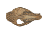
The present 3D Dataset contains the 3D model of a specimen of Metamynodon planifrons (UNISTRA.2015.0.1106) described and figured in: Veine-Tonizzo, L., Tissier, J., Bukhsianidze, M., Vasilyan, D., Becker, D., 2023, Cranial morphology and phylogenetic relationships of Amynodontidae Scott & Osborn, 1883 (Perissodactyla, Rhinocerotoidea).
Metamynodon planifrons UNISTRA.2015.0.1106 View specimen
|
M3#716Textured 3D surface model of the skull of the specimen UNISTRA.2015.0.1106 with right C1 and both rows of P2-M3. Type: "3D_surfaces"doi: 10.18563/m3.sf.716 state:published |
Download 3D surface file |
This contribution contains the 3D models of the ossicles of a protocetid archaeocete from the locality of Kpogamé, Togo, described and figured in the publication of Mourlam and Orliac (2019).
indet. indet. UM KPG-M 73 View specimen

|
M3#407stapes Type: "3D_surfaces"doi: 10.18563/m3.sf.407 state:published |
Download 3D surface file |

|
M3#408Incus Type: "3D_surfaces"doi: 10.18563/m3.sf.408 state:published |
Download 3D surface file |

|
M3#409Malleus Type: "3D_surfaces"doi: 10.18563/m3.sf.409 state:published |
Download 3D surface file |
In this contribution, we describe the external and internal morphology of a delphinid petrosal bone collected from Ahu Tahai, a burial site located on the Southwestern coast of Easter Island, at Hangaroa. We discuss the taxonomic attribution of this archaeological item and describe its internal structures based on µCT data, including the bony labyrinth and the nerve and vein patterns. Identification of the nerves exists lead us to relocate the identification of the foramen singulare in delphinid petrosals.
indet. indet. AT1 View specimen
|
M3#420Stapes Type: "3D_surfaces"doi: 10.18563/m3.sf.420 state:published |
Download 3D surface file |
|
M3#421petrosal bone Type: "3D_surfaces"doi: 10.18563/m3.sf.421 state:published |
Download 3D surface file |
|
M3#422in situ bony labyrinth Type: "3D_surfaces"doi: 10.18563/m3.sf.422 state:published |
Download 3D surface file |
|
M3#423bony labyrinth and associated nerves and blood vessels Type: "3D_surfaces"doi: 10.18563/m3.sf.423 state:published |
Download 3D surface file |
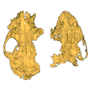
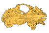
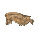
This contribution provides for the first time the 3D model of the type specimen of Molassitherium delemontense (Mammalia, Rhinocerotidae) described in the following publication: Becker et al. (2013), Journal of Systematic Palaeontology, Vol. 11, Issue 8, 947–972, https://doi.org/10.1080/14772019.2012.699007. Conservation issues of the specimen and solutions using 3D model and 3D prints are detailed.
Molassitherium delemontense MJSN POI007–245 View specimen
|
M3#384Skull of Molassitherium delemontense Becker and Antoine, 2013 (in Becker et al. 2013): holotype Type: "3D_surfaces"doi: 10.18563/m3.sf.384 state:published |
Download 3D surface file |
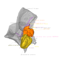
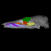
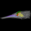
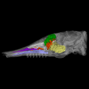
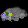
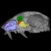
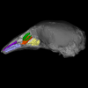
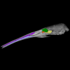
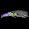
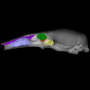
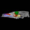
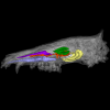
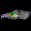
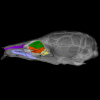
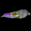
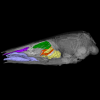
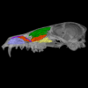
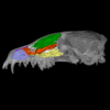
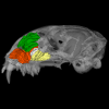
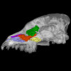
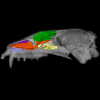
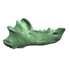
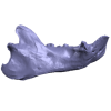
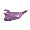
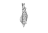
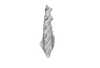
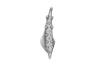
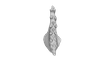
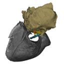
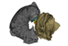
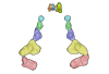
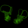
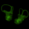
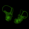
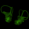
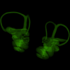
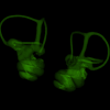
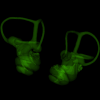
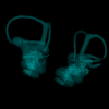
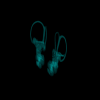
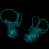
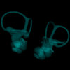
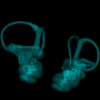
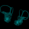
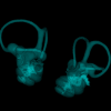
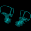
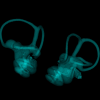
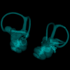
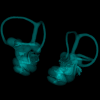
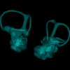
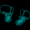
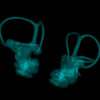
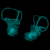
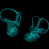
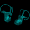
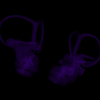
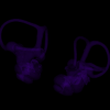
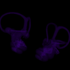
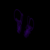
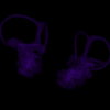
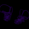
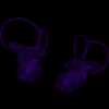
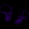
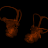
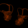
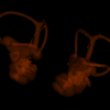
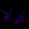
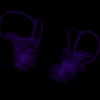
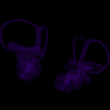
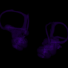
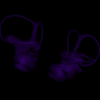
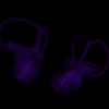
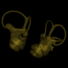
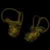
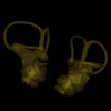
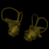
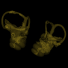
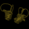
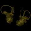
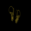
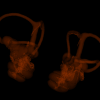
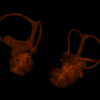
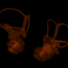
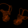
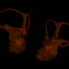
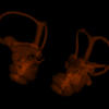
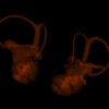
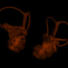
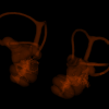
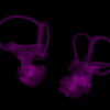
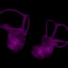
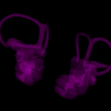
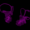
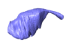
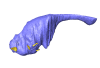
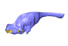
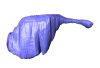
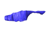
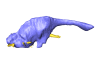
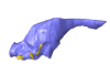
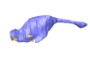
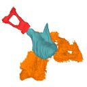
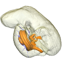
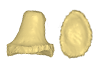
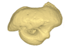
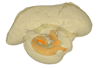
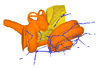
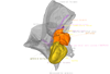
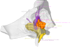
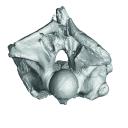
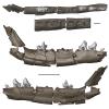
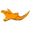
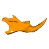
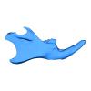
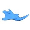
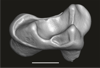
This project presents the osteological connexions of the petrosal bone of the extant Hippopotamidae Hippopotamus amphibius and Choeropsis liberiensis by a virtual osteological dissection of the ear region. The petrosal, the bulla, the sinuses and the major morphological features surrounding the petrosal bone are labelled, both in situ and in an exploded model presenting disassembly views. The directional underwater hearing mode of Hippopotamidae is discussed based on the new observations.
Choeropsis liberiensis UPPal-M09-5-005a View specimen
|
M3#1Labelled compact model of the right ear region of Choeropsis liberiensis (UPPal-M09-5-005a) Type: "3D_surfaces"doi: 10.18563/m3.sf1 state:published |
Download 3D surface file |

|
M3#2Labelled exploded model of the right ear region of Choeropsis liberiensis (UPPal-M09-5-005a) Type: "3D_surfaces"doi: 10.18563/m3.sf2 state:published |
Download 3D surface file |
Hippopotamus amphibius UM N179 View specimen
|
M3#3Labelled compact model of the right ear region of Hippopotamus amphibius (UM N 179) Type: "3D_surfaces"doi: 10.18563/m3.sf3 state:published |
Download 3D surface file |

|
M3#4Labelled exploded model of the right ear region of Hippopotamus amphibius (UM N 179) Type: "3D_surfaces"doi: 10.18563/m3.sf4 state:published |
Download 3D surface file |
The present 3D Dataset contains the 3D model analyzed in Wazir, W. A., Sehgal, R. K., Čerňanský, A., Patnaik, R., Kumar, N., Singh, A. P. and Singh, N. P. 2022. A find from the Ladakh Himalaya reveals a survival of madtsoiid snakes (Serpentes, Madtsoiidae) in India through the late Oligocene. Journal of Vertebrate Paleontology, 41(6), e2058401. https://doi.org/10.1080/02724634.2021.2058401
indet. indet. WIMF/A 4816 View specimen

|
M3#1754Vertebra Type: "3D_surfaces"doi: 10.18563/m3.sf.1754 state:published |
Download 3D surface file |
This contribution contains the three-dimensional models of the most informative fossil material attributed to both Peratherium musivum Gernelle, 2024, and Peratherium maximum (Crochet, 1979), respectively from early and middle early Eocene French localities. These specimens, which document the emergence of the relatively large peratheriines, were analyzed and discussed in: Gernelle et al. (2024), Dental morphology evolution in early peratheriines, including a new morphologically cryptic species and findings on the largest early Eocene European metatherian. https://doi.org/10.1080/08912963.2024.2403602
Peratherium musivum MNHN.F.SN122 View specimen
|
M3#16403D surface model of MNHN.F.SN122, right M3 Type: "3D_surfaces"doi: 10.18563/m3.sf.1640 state:published |
Download 3D surface file |
Peratherium musivum MNHN.F.RI220 View specimen
|
M3#16413D surface model of MNHN.F.RI220, left M2 (partial) Type: "3D_surfaces"doi: 10.18563/m3.sf.1641 state:published |
Download 3D surface file |
Peratherium musivum MNHN.F.RI296 View specimen
|
M3#16423D surface model of MNHN.F.RI296, right M1 (partial) Type: "3D_surfaces"doi: 10.18563/m3.sf.1642 state:published |
Download 3D surface file |
Peratherium musivum MNHN.F.RI368 View specimen
|
M3#16433D surface model of MNHN.F.RI368, right m2 Type: "3D_surfaces"doi: 10.18563/m3.sf.1643 state:published |
Download 3D surface file |
Peratherium musivum MNHN.F.RI385 View specimen
|
M3#16443D surface model of MNHN.F.RI385, left m1 Type: "3D_surfaces"doi: 10.18563/m3.sf.1644 state:published |
Download 3D surface file |
Peratherium maximum UM-BRI-17 View specimen
|
M3#16453D surface model of UM-BRI-17, right hemi-mandible with p1-p3, m1-m3 alveoli, and m4 Type: "3D_surfaces"doi: 10.18563/m3.sf.1645 state:published |
Download 3D surface file |
This contribution contains 3D models of mandibles of Cypriot mice (Mus cypriacus) and house mice (Mus musculus domesticus) from the island of Cyprus. The niche partitioning of the two species was investigated using isotopic ecology, geometric morphometrics and biomechanics. Both species displayed generalist feeding behavior, modulated by fine-tuned adaptation to their feeding habits. The house mouse mandible, with a relatively large masseter area and an optimization for incisor biting, appears as an all-rounder tool for foraging on diverse non-natural items.
These models are analyzed in the following publication: Renaud et al 2024, “Trophic differentiation between the endemic Cypriot mouse and the house mouse: a study coupling stable isotopes and morphometrics”, https://doi.org/10.1007/s10914-024-09740-5
Mus cypriacus Cypriacus_5GE View specimen
|
M3#15843D model of the right mandible Type: "3D_surfaces"doi: 10.18563/m3.sf.1584 state:published |
Download 3D surface file |
Mus cypriacus Cypriacus_BET2 View specimen

|
M3#15853D model of the right mandible Type: "3D_surfaces"doi: 10.18563/m3.sf.1585 state:published |
Download 3D surface file |
Mus cypriacus Cypriacus_FON1 View specimen
|
M3#15863D model of the right mandible Type: "3D_surfaces"doi: 10.18563/m3.sf.1586 state:published |
Download 3D surface file |
Mus cypriacus Cypriacus_FON2 View specimen
|
M3#15873D model of the right mandible Type: "3D_surfaces"doi: 10.18563/m3.sf.1587 state:published |
Download 3D surface file |
Mus cypriacus Cypriacus_KOU1 View specimen
|
M3#15883D model of the right mandible Type: "3D_surfaces"doi: 10.18563/m3.sf.1588 state:published |
Download 3D surface file |
Mus musculus Cyprus_dom_KOF1 View specimen
|
M3#15893D model of the right mandible Type: "3D_surfaces"doi: 10.18563/m3.sf.1589 state:published |
Download 3D surface file |
Mus musculus Cyprus_dom_LEF1 View specimen
|
M3#15903D model of the right mandible Type: "3D_surfaces"doi: 10.18563/m3.sf.1590 state:published |
Download 3D surface file |
Mus musculus Cyprus_dom_MEN1 View specimen

|
M3#15913D model of the right mandible Type: "3D_surfaces"doi: 10.18563/m3.sf.1591 state:published |
Download 3D surface file |
Mus musculus Cyprus_dom_TSE2 View specimen
|
M3#15923D model of the mirrored left mandible Type: "3D_surfaces"doi: 10.18563/m3.sf.1592 state:published |
Download 3D surface file |
Mus musculus Cyprus_dom_XYL5 View specimen
|
M3#15933D model of the right mandible Type: "3D_surfaces"doi: 10.18563/m3.sf.1593 state:published |
Download 3D surface file |

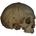
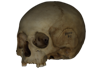
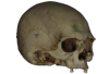
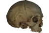
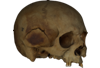
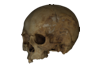
The present 3D Dataset contains the 3D model analyzed in the publication : On Roth’s “human fossil” from Baradero, Buenos Aires Province, Argentina: morphological and genetic analysis. The “human fossil” from Baradero, Buenos Aires Province, Argentina, is a collection of skeleton parts first recovered by Swiss paleontologist Santiago Roth and further studied by anthropologist Rudolf Martin. By the end of the 19th century and beginning of the 20th century it was considered as one of the oldest human skeletons from the southern cone. We studied the cranial anatomy and contextualized the ancient individual remains. We discuss the context of the finding, conducted an osteobiographical assessment and performed a 3D virtual reconstruction of the skull, using micro-CT-scans on selected skull fragments and the mandible. This was followed by the extraction of bone tissue and teeth samples for radiocarbon and genetic analyses, which brought only limited results due to poor preservation and possible contamination. We estimate that the individual from Baradero is a middle-aged adult male. We conclude that the revision of foundational collections with current methodological tools brings new insights and clarifies long held assumptions on the significance of samples that were recovered when archaeology was not yet professionalized.
Homo sapiens PIMUZ A/V 4217 View specimen
|
M3#11983D virtual reconstruction of the skull Type: "3D_surfaces"doi: 10.18563/m3.sf.1198 state:published |
Download 3D surface file |
The present Dataset contains the micro-CT scan of the head of an anonymous 54 year old female donor, at a voxel resolution of 145µm. The skin of the face has been masked in order to avoid the donor to be recognized.
Homo sapiens UM_HS_2018_09_13 View specimen
|
M3#1152Micro-ct data set Type: "3D_CT"doi: 10.18563/m3.sf.1152 state:published |
Download CT data |

This contribution contains the three-dimensional digital model of one isolated fossil tooth of an anthropoid primate (Ashaninkacebus simpsoni), discovered in sedimentary deposits located on the upper Rio Juruá in State of Acre, Brazil (Western Amazonia). This fossil was described, figured and discussed in the following publication: Marivaux et al. (2023), An eosimiid primate of South Asian affinities in the Paleogene of Western Amazonia and the origin of New World monkeys. Proceedings of the National Academy of Sciences USA. https://doi.org/10.1073/pnas.2301338120
Ashaninkacebus simpsoni UFAC-CS 066 View specimen
|
M3#1114Right first upper molar (rM1), pristine. Type: "3D_surfaces"doi: 10.18563/m3.sf.1114 state:published |
Download 3D surface file |


