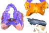
Explodable 3D Dog Skull for Veterinary Education
3D models of Ocnotherium skull
3D models of Kalakocetus, the earliest Cetacea
3D GM dataset of bird skeletal variation
Skeletal embryonic development in the catshark
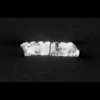
Bony connexions of the petrosal bone of extant hippos
bony labyrinth (14) , inner ear (11) , geometric morphometrics (10) , CT-scan (10) , Eocene (10) , Micro-CT (9) , Miocene (8)
Lionel Hautier (24) , Maëva Judith Orliac (23) , Laurent Marivaux (18) , Renaud Lebrun (14) , Rodolphe Tabuce (14) , Pierre-Olivier Antoine (13) , Bastien Mennecart (13)
|
3D data and models related to the publication: An updated description of the osteology of the pancake tortoise Malacochersus tornieri (Testudines: Testudinidae) with special focus on intraspecific variation.Anna-Katharina Mautner
Published online: 25/01/2017 |
|
M3#129Virtual brain and inner ear endocast of Malacochersus tornieri (ZM 100.102; Zoological Museum of The University of Zurich). This virtual model is accompanied by the 3D dataset. Blue, endocranium; red, blood vessels; purple, semicircular canals; yellow, cranial nerves. Type: "3D_surfaces"doi: 10.18563/m3.sf.129 state:published |
Download 3D surface file |
|
M3#1303D dataset of skull of Malacochersus tornieri (ZM 100.102) Type: "3D_CT"doi: 10.18563/m3.sf.130 state:published |
Download CT data |
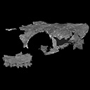
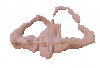
The present 3D Dataset contains the 3D models analyzed in Costeur L., Mennecart B., Müller B., Schulz G., 2016. Prenatal growth stages show the development of the ruminant bony labyrinth and petrosal bone. Journal of Anatomy. https://doi.org/10.1111/joa.12549
Bos taurus NMB3038 View specimen
|
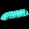
M3#124Right bony labyrinth of a Bos taurus foetus (gestational age 115 days) Type: "3D_surfaces"doi: 10.18563/m3.sf.124 state:published |
Download 3D surface file |
Bos taurus NMB3367 View specimen
|
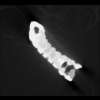
M3#125Right bony labyrinth of a Bos taurus foetus (gestational age 165 days) Type: "3D_surfaces"doi: 10.18563/m3.sf.125 state:published |
Download 3D surface file |
Bos taurus NMB3365 View specimen
|
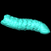
M3#126Right bony labyrinth of a Bos taurus foetus (gestational age 210 days) Type: "3D_surfaces"doi: 10.18563/m3.sf.126 state:published |
Download 3D surface file |
Bos taurus NMB2855 View specimen
|
M3#127Right bony labyrinth of a Bos taurus foetus (gestational age 260 days) Type: "3D_surfaces"doi: 10.18563/m3.sf.127 state:published |
Download 3D surface file |
Bos taurus NMB1037 View specimen
|
M3#128Left bony labyrinth of an adult Bos taurus Type: "3D_surfaces"doi: 10.18563/m3.sf.128 state:published |
Download 3D surface file |
This contribution contains the 3D models described and figured in the following publication: Molnar, JL, Pierce, SE, Bhullar, B-A, Turner, AH, Hutchinson, JR (accepted). Morphological and functional changes in the crocodylomorph vertebral column with increasing aquatic adaptation. Royal Society Open Science.
Protosuchus richardsoni AMNH-VP 3024 View specimen

|
M3#448th and 9th dorsal vertebrae, 1st and 2nd lumbar vertebrae, and 5th lumbar and sacral vertebrae. Type: "3D_surfaces"doi: 10.18563/m3.sf44 state:published |
Download 3D surface file |
Terrestrisuchus gracilis NHM-PV R 7562 View specimen

|
M3#451st and 2nd lumbar vertebrae, and 5th lumbar and sacral vertebrae Type: "3D_surfaces"doi: 10.18563/m3.sf45 state:published |
Download 3D surface file |
Pelagosaurus typus NHM-PV OR 32598 View specimen

|
M3#467th and 8th dorsal vertebrae, 11th and 12th dorsal vertebrae, 15th dorsal vertebra and sacral vertebra. Type: "3D_surfaces"doi: 10.18563/m3.sf46 state:published |
Download 3D surface file |
Metriorhynchus superciliosus NHM-PV R 2054 View specimen

|
M3#476th and 7th dorsal vertebrae, 10th and 11th dorsal vertebrae, 17th dorsal vertebra and sacral vertebra Type: "3D_surfaces"doi: 10.18563/m3.sf47 state:published |
Download 3D surface file |
Crocodylus niloticus FNC0 View specimen

|
M3#487th and 8th dorsal vertebrae, 1st and 2nd lumbar vertebrae, 5th lumbar vertebra and sacral vertebra. Type: "3D_surfaces"doi: 10.18563/m3.sf48 state:published |
Download 3D surface file |
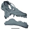
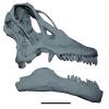
The study of titanosaur paleobiology has been severely hampered by the incomplete nature of their fossil record, particularly the scarcity of well-preserved and relatively complete cranial remains. Even the most complete titanosaur skulls are often fractured, incomplete, or deformed, which has resulted in a limited knowledge of the paleobiology related to cranial anatomy, especially functional morphology. In this context, we present the digital restoration of the skull of the Argentinean titanosaur Sarmientosaurus musacchioi, created using the open-source 3D modeling software Blender. The digitally restored model is freely accessible to other researchers, facilitating broader research and comparative studies.
Sarmientosaurus mussacchioi MDT-PV 02 View specimen
|
M3#1594Cranium and mandible of Sarmientosaurus mussacchioi Type: "3D_surfaces"doi: 10.18563/m3.sf.1594 state:published |
Download 3D surface file |

|
M3#1599Original object provided by Gabriel Casal (cranium) Type: "3D_surfaces"doi: 10.18563/m3.sf.1599 state:published |
Download 3D surface file |
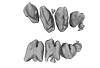
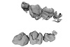
The present contribution contains the 3D models of fossil humeri and ilia of anurans from various Eocene-Miocene deposits of Peruvian Amazonia. These fossils were described and figured in the following publication: Jansen et al. (2023), First Eocene–Miocene anuran fossils from Peruvian Amazonia: insights into Neotropical frog evolution and diversity. Papers in Palaeontology, The Palaeontological Association.
Indet. indet. MUSM 4746 View specimen
|
M3#1231Humeral fragment (distal end) Type: "3D_surfaces"doi: 10.18563/m3.sf.1231 state:published |
Download 3D surface file |
Indet. indet. MUSM 4747 View specimen
|
M3#1232Humeral fragment (distal end) Type: "3D_surfaces"doi: 10.18563/m3.sf.1232 state:published |
Download 3D surface file |
Indet. indet. MUSM 4748 View specimen
|
M3#1233Humeral fragment (distal end) Type: "3D_surfaces"doi: 10.18563/m3.sf.1233 state:published |
Download 3D surface file |
Indet. indet. MUSM 4755 View specimen
|
M3#1234Humeral fragment (distal end) Type: "3D_surfaces"doi: 10.18563/m3.sf.1234 state:published |
Download 3D surface file |
Indet. indet. MUSM 4756 View specimen
|
M3#1235Humeral fragment (distal end) Type: "3D_surfaces"doi: 10.18563/m3.sf.1235 state:published |
Download 3D surface file |
Indet. indet. MUSM 4757 View specimen
|
M3#1236Humeral fragment (distal end) Type: "3D_surfaces"doi: 10.18563/m3.sf.1236 state:published |
Download 3D surface file |
Indet. indet. MUSM 4761 View specimen
|
M3#1237Humeral fragment (distal end) Type: "3D_surfaces"doi: 10.18563/m3.sf.1237 state:published |
Download 3D surface file |
Indet. indet. MUSM 4763 View specimen
|
M3#1238Humeral fragment (distal end) Type: "3D_surfaces"doi: 10.18563/m3.sf.1238 state:published |
Download 3D surface file |
Indet. indet. MUSM 4765 View specimen
|
M3#1239Humeral fragment (distal end) Type: "3D_surfaces"doi: 10.18563/m3.sf.1239 state:published |
Download 3D surface file |
Indet. indet. MUSM 4766 View specimen
|
M3#1240Humeral fragment (distal end) Type: "3D_surfaces"doi: 10.18563/m3.sf.1240 state:published |
Download 3D surface file |
Indet. indet. MUSM 4775 View specimen
|
M3#1241Humeral fragment (distal end) Type: "3D_surfaces"doi: 10.18563/m3.sf.1241 state:published |
Download 3D surface file |
cf. Pipa sp. MUSM 4776 View specimen
|
M3#1242Humeral fragment (distal end) Type: "3D_surfaces"doi: 10.18563/m3.sf.1242 state:published |
Download 3D surface file |
Indet. indet. MUSM 4788 View specimen
|
M3#1243Ilial fragment Type: "3D_surfaces"doi: 10.18563/m3.sf.1243 state:published |
Download 3D surface file |
Indet. indet. MUSM 4789 View specimen
|
M3#1244Ilial fragment Type: "3D_surfaces"doi: 10.18563/m3.sf.1244 state:published |
Download 3D surface file |
Indet. indet. MUSM 4790 View specimen
|
M3#1245Ilial fragment Type: "3D_surfaces"doi: 10.18563/m3.sf.1245 state:published |
Download 3D surface file |
Indet. indet. MUSM 4792 View specimen
|
M3#1246Ilial fragment Type: "3D_surfaces"doi: 10.18563/m3.sf.1246 state:published |
Download 3D surface file |
Indet. indet. MUSM 4793 View specimen
|
M3#1247Ilial fragment Type: "3D_surfaces"doi: 10.18563/m3.sf.1247 state:published |
Download 3D surface file |
Indet. indet. MUSM 4794 View specimen
|
M3#1249Ilial fragment Type: "3D_surfaces"doi: 10.18563/m3.sf.1249 state:published |
Download 3D surface file |
Indet. indet. MUSM 4795 View specimen
|
M3#1250Ilial fragment Type: "3D_surfaces"doi: 10.18563/m3.sf.1250 state:published |
Download 3D surface file |
cf. Pipa sp. MUSM 4796 View specimen
|
M3#1251Ilial fragment Type: "3D_surfaces"doi: 10.18563/m3.sf.1251 state:published |
Download 3D surface file |
cf. Pipa sp. MUSM 4797 View specimen
|
M3#1252Ilial fragment Type: "3D_surfaces"doi: 10.18563/m3.sf.1252 state:published |
Download 3D surface file |
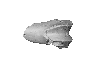
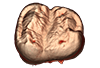
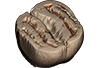
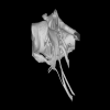
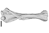
Turtles are one of the most impressive vertebrates. Much of the body is either hidden in a shell or can be drawn into it. Turtles impress with their individual longevity and their often peaceful disposition. Also, with their resilience, they have survived all extinction events since their emergence in the Late Triassic. Today's diversity of shapes is impressive and ranges from the large and high domed Galapagos turtles to the hamster-sized flat pancake turtles. The holotype of one of the oldest fossil turtles, Proganochelys quenstedtii, is housed in the paleontological collection in Tübingen/Germany. Since its discovery some years before 1873, P. quenstedtii has represented the 'prototype' of the turtle and has had an eventful scientific history. It was found in Neuenhaus (Häfner-Neuhausen in Schönbuch forest), Baden-Württemberg, Germany, and stems from Löwenstein-Formation (Weißer Keupersandstein), Late Triassic. The current catalogue number is GPIT-PV-30000. The specimen is listed in the historical inventory “Tübinger Petrefaktenverzeichnis 1841 bis 1896, [folio 326v.]“, as “[catalogue number: PV]16549, Schildkröte Weiser Keupersandstein Hafnerhausen” [turtle from White Keuper Sandstone]. Another, more recent synonym is “GPIT/RE/9396”. The same specimen was presented as uncatalogued by Gaffney (1990). Here we provide a surface scan of the steinkern for easier access of this famous specimen to the scientific community.
Proganochelys quenstedtii GPIT-PV-30000 View specimen
|
M3#967This the surface model of the steinkern of the shell of Proganochelys quenstedtii. Type: "3D_surfaces"doi: 10.18563/m3.sf.967 state:published |
Download 3D surface file |
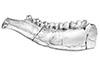
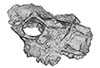
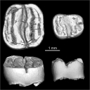
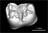
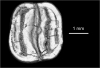
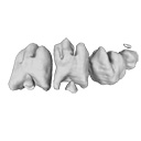
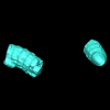
The present 3D Dataset contains the 3D models of the holotype mandible and referred fragmented skull of the new species Amphimoschus xishuiensis analyzed in the article Li, Y.-K., Mennecart, B., Aiglstorfer, M., Ni, X.-J., Li, Q., Deng, T. 2021. The early evolution of cranial appendages in Bovoidea revealed by new species of Amphimoschus (Mammalia: Ruminantia) from China. Zoological Journal of the Linnean Society https://doi.org/10.1093/zoolinnean/zlab053
Amphimoschus xishuiensis IVPP V 25521.1 View specimen
|
M3#803the holotype, a right hemimandible with tooth row p2 to m3 Type: "3D_surfaces"doi: 10.18563/m3.sf.803 state:published |
Download 3D surface file |
Amphimoschus xishuiensis IVPP V 25521.2 View specimen
|
M3#804referred material, anterior part of a skull with right P4-M3 and left P3-M2 Type: "3D_surfaces"doi: 10.18563/m3.sf.804 state:published |
Download 3D surface file |
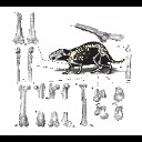
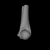
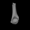
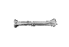
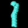
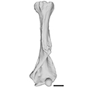
This contribution contains the 3D models of postcranial bones (humerus, ulna, innominate, femur, tibia, astragalus, navicular, and metatarsal III) described and figured in the following publication: “Postcranial morphology of the extinct rodent Neoepiblema (Rodentia: Chinchilloidea): insights into the paleobiology of neoepiblemids”.
Neoepiblema acreensis UFAC 3549 View specimen
|
M3#719UFAC 3549, left humerus missing the proximal region. Type: "3D_surfaces"doi: 10.18563/m3.sf.719 state:published |
Download 3D surface file |
Neoepiblema acreensis UFAC 5076 View specimen
|
M3#720UFAC 5076, right humerus missing the proximal region. Type: "3D_surfaces"doi: 10.18563/m3.sf.720 state:published |
Download 3D surface file |
Neoepiblema acreensis UFAC 1939 View specimen
|
M3#721UFAC 1939, right ulna missing the olecranon epiphysis and the distal region. Type: "3D_surfaces"doi: 10.18563/m3.sf.721 state:published |
Download 3D surface file |
Neoepiblema acreensis UFAC 3697 View specimen
|
M3#722UFAC 3697, right innominate bone. Type: "3D_surfaces"doi: 10.18563/m3.sf.722 state:published |
Download 3D surface file |
Neoepiblema acreensis UFAC 2574 View specimen
|
M3#723UFAC 2574, proximal region of a left femur. Type: "3D_surfaces"doi: 10.18563/m3.sf.723 state:published |
Download 3D surface file |
Neoepiblema acreensis UFAC 2937 View specimen
|
M3#724UFAC 2937, right femur with damaged proximal region. Type: "3D_surfaces"doi: 10.18563/m3.sf.724 state:published |
Download 3D surface file |
Neoepiblema acreensis UFAC 2210 View specimen
|
M3#725UFAC 2210, distal region of a right femur. Type: "3D_surfaces"doi: 10.18563/m3.sf.725 state:published |
Download 3D surface file |
Neoepiblema acreensis UFAC 1887 View specimen
|
M3#726UFAC 1887, right tibia Type: "3D_surfaces"doi: 10.18563/m3.sf.726 state:published |
Download 3D surface file |
Neoepiblema acreensis UFAC 1840 View specimen
|
M3#727UFAC 1840, left astragalus. Type: "3D_surfaces"doi: 10.18563/m3.sf.727 state:published |
Download 3D surface file |
Neoepiblema acreensis UFAC 2549 View specimen
|
M3#728UFAC 2549, right astragalus. Type: "3D_surfaces"doi: 10.18563/m3.sf.728 state:published |
Download 3D surface file |
Neoepiblema acreensis UFAC 3672 View specimen
|
M3#729UFAC 3672, right navicular. Type: "3D_surfaces"doi: 10.18563/m3.sf.729 state:published |
Download 3D surface file |
Neoepiblema acreensis UFAC 2116 View specimen
|
M3#730UFAC 2116, left metatarsal III. Type: "3D_surfaces"doi: 10.18563/m3.sf.730 state:published |
Download 3D surface file |
Neoepiblema horridula UFAC 3260 View specimen

|
M3#731UFAC 3260, fragmented left innominate. Type: "3D_surfaces"doi: 10.18563/m3.sf.731 state:published |
Download 3D surface file |
Neoepiblema horridula UFAC 2620 View specimen
|
M3#732UFAC 2620, distal region of a right femur. Type: "3D_surfaces"doi: 10.18563/m3.sf.732 state:published |
Download 3D surface file |
Neoepiblema horridula UFAC 2737 View specimen
|
M3#733UFAC 2737, proximal region of right femur. Type: "3D_surfaces"doi: 10.18563/m3.sf.733 state:published |
Download 3D surface file |
Neoepiblema horridula UFAC 3202 View specimen
|
M3#734UFAC 3202, right tibia, missing the proximalmost and distal portions. Type: "3D_surfaces"doi: 10.18563/m3.sf.734 state:published |
Download 3D surface file |
Neoepiblema horridula UFAC 3212 View specimen
|
M3#735UFAC 3212, left astragalus. Type: "3D_surfaces"doi: 10.18563/m3.sf.735 state:published |
Download 3D surface file |
This contribution provides the raw files for the μCT-scan data and renderings of the three-dimensional digital models of two fossil teeth of a geomyin geomorph rodent (Caribeomys merzeraudi), discovered from lower Oligocene deposits of Puerto Rico, San Sebastian Formation (locality LACM Loc. 8060). These fossils were described, figured and discussed in the following publication: Marivaux et al. (2021), An unpredicted ancient colonization of the West Indies by North American rodents: dental evidence of a geomorph from the early Oligocene of Puerto Rico. Papers in Palaeontology. https://doi.org/10.1002/spp2.1388
Caribeomys merzeraudi LACM 162478 View specimen
|
M3#712Right lower dp4: isolated deciduous premolar. The specimen was scanned with a resolution of 5 µm using a μ-CT-scanning station EasyTom 150 / Rx Solutions (Montpellier RIO Imaging, ISE-M, Montpellier, France). AVIZO 7.1 (Visualization Sciences Group) software was used for visualization, segmentation, and 3D rendering. This isolated tooth was prepared within a “labelfield” module of AVIZO, using the segmentation threshold selection tool. Type: "3D_surfaces"doi: 10.18563/m3.sf.712 state:published |
Download 3D surface file |
|
M3#7145µm µCT data set . Right lower dp4: isolated deciduous premolar. The specimen was scanned with a resolution of 5 µm using a μ-CT-scanning station EasyTom 150 / Rx Solutions (Montpellier RIO Imaging, ISE-M, Montpellier, France). Type: "3D_CT"doi: 10.18563/m3.sf.714 state:published |
Download CT data |
Caribeomys merzeraudi LACM 162449 View specimen
|
M3#713Right lower molar (m1 or m2). The specimen was scanned with a resolution of 4.5 µm using a μ-CT-scanning station EasyTom 150 / Rx Solutions (Montpellier RIO Imaging, ISE-M, Montpellier, France). AVIZO 7.1 (Visualization Sciences Group) software was used for visualization, segmentation, and 3D rendering. This isolated tooth was prepared within a “labelfield” module of AVIZO, using the segmentation threshold selection tool. Type: "3D_surfaces"doi: 10.18563/m3.sf.713 state:published |
Download 3D surface file |
|
M3#715µCT data at 4.5µm Type: "3D_CT"doi: 10.18563/m3.sf.715 state:published |
Download CT data |
The present 3D Dataset contains the 3D model of the brain endocast of Neoepiblema acreensis analyzed in “Small within the largest: Brain size and anatomy of the extinct Neoepiblema acreensis, a giant rodent from the Neotropics”. The 3D model was generated using CT-Scanning and techniques of virtual reconstruction.
Neoepiblema acreensis UFAC 4515 View specimen
|
M3#502Brain endocast of Neoepiblema acreensis Type: "3D_surfaces"doi: 10.18563/m3.sf.502 state:published |
Download 3D surface file |
The present 3D Dataset contains the 3D models analyzed in ”Morphological features of tooth development and replacement in the rabbit Oryctolagus cuniculus”, Archives of Oral Biology, https://doi.org/10.1016/j.archoralbio.2019.104576
Oryctogalus cuniculus E14 View specimen
|
M3#390Right cheek teeth, Left and right incisors at 14 dpf Type: "3D_surfaces"doi: 10.18563/m3.sf.390 state:published |
Download 3D surface file |
Oryctogalus cuniculus E16 View specimen
|
M3#391Left cheek teeth, Left and right incisors at 16 dpf Type: "3D_surfaces"doi: 10.18563/m3.sf.391 state:published |
Download 3D surface file |
Oryctogalus cuniculus E18 View specimen
|
M3#392Left cheek teeth and incisors at 18 dpf Type: "3D_surfaces"doi: 10.18563/m3.sf.392 state:published |
Download 3D surface file |
Oryctogalus cuniculus E20 View specimen
|
M3#393Left cheek teeth and incisors at 20 dpf Type: "3D_surfaces"doi: 10.18563/m3.sf.393 state:published |
Download 3D surface file |
Oryctogalus cuniculus E22 View specimen
|
M3#394Left lower cheek teeth and incisors, right upper cheek teeth and incisors at 22 dpf Type: "3D_surfaces"doi: 10.18563/m3.sf.394 state:published |
Download 3D surface file |
Oryctogalus cuniculus E24 View specimen
|
M3#395Left cheek teeth and incisors at 24 dpf Type: "3D_surfaces"doi: 10.18563/m3.sf.395 state:published |
Download 3D surface file |
Oryctogalus cuniculus E28 View specimen
|
M3#396Right cheek teeth and incisors at 28 dpf Type: "3D_surfaces"doi: 10.18563/m3.sf.396 state:published |
Download 3D surface file |
Oryctogalus cuniculus E26 View specimen
|
M3#397Right cheek teeth and incisors at 26 dpf Type: "3D_surfaces"doi: 10.18563/m3.sf.397 state:published |
Download 3D surface file |
The present 3D Dataset contains the 3D model analyzed in the article : Dubied et al. (2021), Endocranium and ecology of Eurotherium theriodis, a European hyaenodont mammal from the Lutetian. Acta Palaeontologica Polonica 2021, https://doi.org/10.4202/app.00771.2020
Eurotherium theriodis NMB.Em12 View specimen
|
M3#381NMB.Em12 unprepared specimen Type: "3D_surfaces"doi: 10.18563/m3.sf.381 state:published |
Download 3D surface file |
|
M3#382NMB.Em12 cranium Type: "3D_surfaces"doi: 10.18563/m3.sf.382 state:published |
Download 3D surface file |
|
M3#383NMB.Em12 endocast Type: "3D_surfaces"doi: 10.18563/m3.sf.383 state:published |
Download 3D surface file |
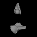
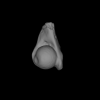
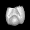
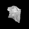
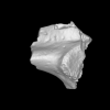
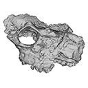
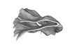
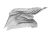
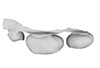
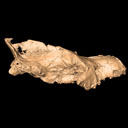
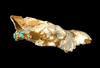
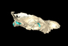
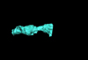
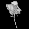
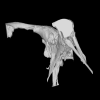
This contribution contains the 3D models described and figured in the following publication: Mennecart B., de Perthuis Ad., Rössner G.E., Guzmán J.A., de Perthuis Au., Costeur L. The first French tragulid skull (Mammalia, Ruminantia, Tragulidae) and associated tragulid remains from the Middle Miocene of Contres (Loir-et-Cher, France). Comptes Rendus Palévol. https://doi.org/10.1016/j.crpv.2017.08.004
Dorcatherium crassum NMB Fa.213.abg View specimen
|
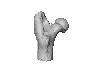
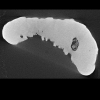
M3#181The 3D surface files of the specimen NMB Fa.213 are the reconstructions of the main skull fragments, the right petrosal bone, and the left bony labyrinth. Type: "3D_surfaces"doi: 10.18563/m3.sf.181 state:published |
Download 3D surface file |

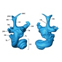
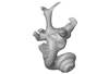
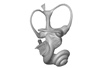
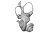
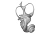
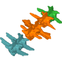
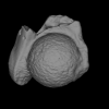
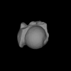
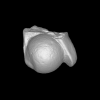
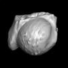
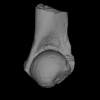
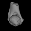
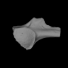
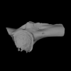
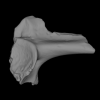
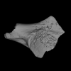
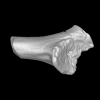
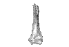
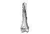
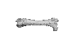
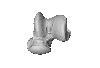
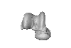
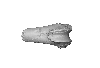
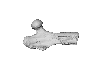
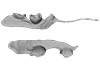
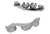
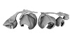
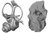
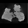
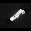
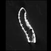
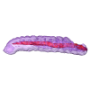
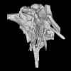
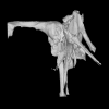
This project presents the 3D models of two isolated petrosals from the Oligocene locality of Pech de Fraysse (Quercy, France) here attributed to the genus Prodremotherium Filhol, 1877. Our aim is to describe the petrosal morphology of this Oligocene “early ruminant” as only few data are available in the literature for Oligocene taxa.
Prodremotherium sp. UM PFY 4053 View specimen

|
M3#7Labelled 3D model of right isolated petrosal of Prodremotherium sp. from Pech de Fraysse (Quercy, MP 28) Type: "3D_surfaces"doi: 10.18563/m3.sf7 state:published |
Download 3D surface file |
Prodremotherium sp. UM PFY 4054 View specimen

|
M3#8Labelled 3D model of right isolated petrosal of Prodremotherium sp. from Pech de Fraysse (Quercy, MP 28) Type: "3D_surfaces"doi: 10.18563/m3.sf8 state:published |
Download 3D surface file |
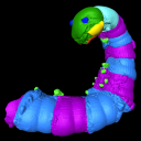
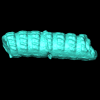
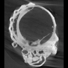
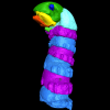
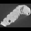
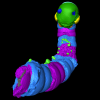
The present 3D Dataset contains the 3D models analyzed in the publication: Mummified Paleogene Spirostreptida and Julida (Arthropoda, Diplopoda) from southern France. Papers in Paleontology.
Protosilvestria sculpta NMB F1935 View specimen
|
M3#1457Paralectotype, 13 diplosegments with the proximal part of the legs Type: "3D_surfaces"doi: 10.18563/m3.sf.1457 state:published |
Download 3D surface file |
|
M3#1657CT data of NMB F1935. Images were reduced by a binning of factor 2. Type: "3D_CT"doi: 10.18563/m3.sf.1657 state:published |
Download CT data |
Protosilvestria sculpta NMB F1936 View specimen
|
M3#1458Lectotype, head with the ten following segments Type: "3D_surfaces"doi: 10.18563/m3.sf.1458 state:published |
Download 3D surface file |
|
M3#1658CT data of NMB F1936. Type: "3D_CT"doi: 10.18563/m3.sf.1658 state:published |
Download CT data |
Protosilvestria sculpta NMB F1937 View specimen
|
M3#1459Paralectotype, seven segments Type: "3D_surfaces"doi: 10.18563/m3.sf.1459 state:published |
Download 3D surface file |
|
M3#1659CT data of NMB F1937 Type: "3D_CT"doi: 10.18563/m3.sf.1659 state:published |
Download CT data |
Protosilvestria sculpta NMB F1938 View specimen
|
M3#1460Paralectotype, ten segments and the telson Type: "3D_surfaces"doi: 10.18563/m3.sf.1460 state:published |
Download 3D surface file |
|
M3#1660CT data of NMB F1938 Type: "3D_CT"doi: 10.18563/m3.sf.1660 state:published |
Download CT data |
Protosilvestria sculpta NMB F1987 View specimen
|
M3#1461Nine segments and the telson Type: "3D_surfaces"doi: 10.18563/m3.sf.1461 state:published |
Download 3D surface file |
|
M3#1661CT data of NMB F1987 Type: "3D_CT"doi: 10.18563/m3.sf.1661 state:published |
Download CT data |
Protosilvestria sculpta NMB F1988 View specimen
|
M3#1462Two parts, first part=telson and four segments, second part=five segments Type: "3D_surfaces"doi: 10.18563/m3.sf.1462 state:published |
Download 3D surface file |
|
M3#1662CT data of NMB F1988 Type: "3D_CT"doi: 10.18563/m3.sf.1662 state:published |
Download CT data |
Protosilvestria sculpta NMB F1989 View specimen
|
M3#1463Telson with 13 segments and the digestive tract Type: "3D_surfaces"doi: 10.18563/m3.sf.1463 state:published |
Download 3D surface file |
|
M3#1663CT data of NMB F1989 Type: "3D_CT"doi: 10.18563/m3.sf.1663 state:published |
Download CT data |
Protosilvestria sculpta NMB F1990 View specimen
|
M3#1464Eight segments and the telson Type: "3D_surfaces"doi: 10.18563/m3.sf.1464 state:published |
Download 3D surface file |
|
M3#1664CT data of NMB F1990 Type: "3D_CT"doi: 10.18563/m3.sf.1664 state:published |
Download CT data |
Protosilvestria sculpta NMB F3743 View specimen
|
M3#1465Seven segments and legs Type: "3D_surfaces"doi: 10.18563/m3.sf.1465 state:published |
Download 3D surface file |
|
M3#1665CT data of NMB F3743. Images were reduced by a binning of factor 2. Type: "3D_CT"doi: 10.18563/m3.sf.1665 state:published |
Download CT data |
Protosilvestria sculpta UM-SND-1704 View specimen
|
M3#1468Head and seven segments Type: "3D_surfaces"doi: 10.18563/m3.sf.1468 state:published |
Download 3D surface file |
|
M3#1666CT data of UM-SND-1704. Images were reduced by a binning of factor 2. Type: "3D_CT"doi: 10.18563/m3.sf.1666 state:published |
Download CT data |
Indet Indet UM-ROQ1-500 View specimen
|
M3#1467Head and ten segments Type: "3D_surfaces"doi: 10.18563/m3.sf.1467 state:published |
Download 3D surface file |
|
M3#1667CT data of UM-ROQ1-500. Images were reduced by a binning of factor 2. Type: "3D_CT"doi: 10.18563/m3.sf.1667 state:published |
Download CT data |
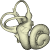
The present 3D Dataset contains 3D models of the cranium surface and of the bony labyrinth endocast of the stem bat Vielasia sigei. They are used by (Hand et al., 2023) to explore the phylogenetic position of this species, to infer its laryngeal echolocating capabilities, and to eventually discuss chiropteran evolution before the crown clade diversification.
Vielasia sigei UM VIE-250 View specimen
|
M3#1269External surface of the cranium Type: "3D_surfaces"doi: 10.18563/m3.sf.1269 state:published |
Download 3D surface file |
|
M3#1270Virtual endocast of the right bony labyrinth Type: "3D_surfaces"doi: 10.18563/m3.sf.1270 state:published |
Download 3D surface file |

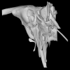
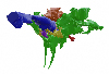
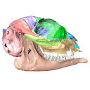
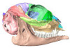
The present 3D Dataset contains the 3D models analyzed in: Perrichon et al., 2023. Neuroanatomy and pneumaticity of Voay robustus and its implications for crocodylid phylogeny and palaeoecology.
Crocodylus niloticus MHNL 50001387 View specimen
|
M3#1202Skull, inner ear, pharyngotympanic sinus and neurovascular system Type: "3D_surfaces"doi: 10.18563/m3.sf.1202 state:published |
Download 3D surface file |
Voay robustus MNHN F.1908-5 View specimen
|
M3#1203Skull, inner ear, pharyngotympanic sinus and neurovascular system Type: "3D_surfaces"doi: 10.18563/m3.sf.1203 state:published |
Download 3D surface file |
Voay robustus NHMUK PV R 36684 View specimen
|
M3#1204Skull, inner ear, pharyngotympanic sinus and neurovascular system Type: "3D_surfaces"doi: 10.18563/m3.sf.1204 state:published |
Download 3D surface file |
Voay robustus NHMUK PV R 36685 View specimen
|
M3#1205Skull, inner ear, pharyngotympanic sinus and neurovascular system Type: "3D_surfaces"doi: 10.18563/m3.sf.1205 state:published |
Download 3D surface file |
Osteolaemus tetraspis UCBLZ 2019-1-236 View specimen
|
M3#1208Skull, inner ear, pharyngotympanic sinus and neurovascular system Type: "3D_surfaces"doi: 10.18563/m3.sf.1208 state:published |
Download 3D surface file |
Mecistops sp. UM N89 View specimen
|
M3#1207Skull, inner ear, pharyngotympanic sinus and neurovascular system Type: "3D_surfaces"doi: 10.18563/m3.sf.1207 state:published |
Download 3D surface file |
Voay robustus NHMUK PV R 2204 View specimen
|
M3#1206Skull, inner ear, pharyngotympanic sinus, intertympanic sinus and neurovascular system Type: "3D_surfaces"doi: 10.18563/m3.sf.1206 state:published |
Download 3D surface file |
The present 3D Dataset contains the 3D model analyzed in the following publication: occurrence of the ground sloth Nothrotheriops (Xenarthra, Folivora) in the Late Pleistocene of Uruguay: New information on its dietary and habitat preferences based on stable isotope analysis. Journal of Mammalian Evolution. https://doi.org/10.1007/s10914-023-09660-w
Nothrotheriops sp. CAV 1466 View specimen
|
M3#1129Left humerus Type: "3D_surfaces"doi: 10.18563/m3.sf.1129 state:published |
Download 3D surface file |
The present 3D Dataset contains the 3D models analyzed in Pochat-Cottilloux Y., Rinder N., Perrichon G., Adrien J., Amiot R., Hua S. & Martin J. E. (2023). The neuroanatomy and pneumaticity of Hamadasuchus from the Cretaceous of Morocco and its significance for the paleoecology of Peirosauridae and other altirostral crocodylomorphs. Journal of Anatomy, https://doi.org/10.1111/joa.13887
Hamadasuchus sp. UCBL-FSL 532408 View specimen
|
M3#10943D volume reconstruction of the braincase osteology Type: "3D_surfaces"doi: 10.18563/m3.sf.1094 state:published |
Download 3D surface file |
|
M3#10963D volume reconstruction of the endocast Type: "3D_surfaces"doi: 10.18563/m3.sf.1096 state:published |
Download 3D surface file |
|
M3#10973D volume reconstruction of the labyrinths Type: "3D_surfaces"doi: 10.18563/m3.sf.1097 state:published |
Download 3D surface file |
|
M3#10983D volume reconstruction of the pneumatic cavities Type: "3D_surfaces"doi: 10.18563/m3.sf.1098 state:published |
Download 3D surface file |
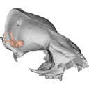
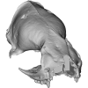
The present 3D Dataset contains the 3D models analyzed in Keppeler, H., Schultz, J. A., Ruf, I., & Martin, T., 2023. Cranial anatomy of Hypisodus minimus (Artiodactyla: Ruminantia) from the Oligocene Brule Formation of North America. Palaeontographica Abteilung A.
Hypisodus minimus SMNK-PAL 27212 View specimen

|
M3#1031CT image stack of a skull of Hypisodus minimus. Also includes a lumbar vertebra and a probable proximal phalanx of digit III or IV. Type: "3D_CT"doi: 10.18563/m3.sf.1031 state:published |
Download CT data |
|
M3#10363D surface models of a skull of Hypisodus minimus (SMNK-PAL27212). The data includes a surface model for: basisphenoid, tympanic bullae, ethmoid (lamina perpendicularis), frontals, jugal (left), jugal (right), lacrimals, lower dentition, mandibles, mastoid processes, maxillaries, maxilloturbinals, nasals, occipital, palatine, parietals, petrosals, presphenoid, squamosals, turbinates, upper dentition, and the vomer. Type: "3D_surfaces"doi: 10.18563/m3.sf.1036 state:published |
Download 3D surface file |
Hypisodus minimus SMNK-PAL 27213 View specimen
|
M3#1033CT image stack of a skull of Hypisodus minimus. Also shows numerous postcranial material including an atlas articulated with the occipital bone, the distal part of a left humerus articulated to radius and ulna, a part of a femur, a part of a tibia and fibula, unidentifiable tarsal bones, parts of the metatarsals II, III, IV and V and their phalanges, a proximal phalanx of digit III or IV, a middle phalanx of digit III or IV, a possible patella and calcaneus, as well as numerous unidentifiable broken bony fragments. Type: "3D_CT"doi: 10.18563/m3.sf.1033 state:published |
Download CT data |
|
M3#10353D surface models of a skull of Hypisodus minimus (SMNK-PAL27213). The data includes a surface model for: atlas, basisphenoid, tympanic bullae, nasals, occipital, the petrosals, and the inner ear. Type: "3D_surfaces"doi: 10.18563/m3.sf.1035 state:published |
Download 3D surface file |


