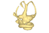
Explodable 3D Dog Skull for Veterinary Education
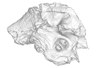
3D models of Ocnotherium skull
3D models of Kalakocetus, the earliest Cetacea
3D GM dataset of bird skeletal variation
Skeletal embryonic development in the catshark
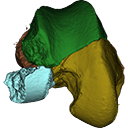
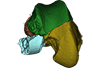
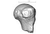
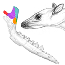
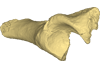
Bony connexions of the petrosal bone of extant hippos
bony labyrinth (14) , inner ear (11) , geometric morphometrics (10) , CT-scan (10) , Eocene (10) , Micro-CT (9) , Miocene (8)
Lionel Hautier (24) , Maëva Judith Orliac (23) , Laurent Marivaux (18) , Renaud Lebrun (14) , Rodolphe Tabuce (14) , Pierre-Olivier Antoine (13) , Bastien Mennecart (13)
|
3D models related to the publication: New remains of Neotropical bunodont litopterns and the systematics of Megadolodinae (Mammalia: Litopterna)Juan D. Carrillo
Published online: 31/08/2023 |
|
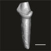
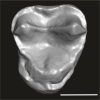
M3#1020Partial mandible with the symphysis and left body, bearing the alveoli of ?i2, right and left ?i3, alveolus of right c and p1, roots of left p1, and the left p2–m3 of Megadolodus molariformes (Litopterna, Mammalia) Type: "3D_surfaces"doi: 10.18563/m3.sf.1020 state:published |
Download 3D surface file |
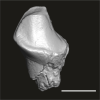
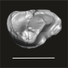
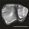
Neodolodus colombianus VPPLT 1696 View specimen
|
M3#1021Almost complete skull with left and right ?I2 and P1–M3 Type: "3D_surfaces"doi: 10.18563/m3.sf.1021 state:published |
Download 3D surface file |
|
M3#1022Partial mandible with complete right and left dentition except for left ?i2 Type: "3D_surfaces"doi: 10.18563/m3.sf.1022 state:published |
Download 3D surface file |
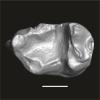
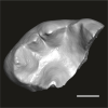
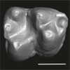
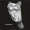
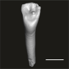
This contribution contains the 3D models of the fossil teeth of two chinchilloid caviomorph rodents (Borikenomys praecursor and Chinchilloidea gen. et sp. indet.) discovered from lower Oligocene deposits of Puerto Rico, San Sebastian Formation (locality LACM Loc. 8060). These fossils were described and figured in the following publication: Marivaux et al. (2020), Early Oligocene chinchilloid caviomorphs from Puerto Rico and the initial rodent colonization of the West Indies. Proceedings of the Royal Society B. http://dx.doi.org/10.1098/rspb.2019.2806
Borikenomys praecursor LACM 162447 View specimen
|
M3#638Right lower m3. This isolated tooth was scanned with a resolution of 6 µm using a μ-CT-scanning station EasyTom 150 / Rx Solutions (Montpellier RIO Imaging, ISE-M, Montpellier, France). AVIZO 7.1 (Visualization Sciences Group) software was used for visualization, segmentation, and 3D rendering. The specimen was prepared within a “labelfield” module of AVIZO, using the segmentation threshold selection tool. Type: "3D_surfaces"doi: 10.18563/m3.sf.638 state:published |
Download 3D surface file |
Borikenomys praecursor LACM 162446 View specimen
|
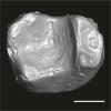
M3#639Fragment of lower molar (most of the mesial part). This isolated broken tooth was scanned with a resolution of 6 µm using a μ-CT-scanning station EasyTom 150 / Rx Solutions (Montpellier RIO Imaging, ISE-M, Montpellier, France). AVIZO 7.1 (Visualization Sciences Group) software was used for visualization, segmentation, and 3D rendering. The specimen was prepared within a “labelfield” module of AVIZO, using the segmentation threshold selection tool. Type: "3D_surfaces"doi: 10.18563/m3.sf.639 state:published |
Download 3D surface file |
indet indet LACM 162448 View specimen
|
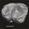
M3#640Fragment of either an upper tooth (mesial laminae) or a lower tooth (distal laminae). The specimen was scanned with a resolution of 6 µm using a μ-CT-scanning station EasyTom 150 / Rx Solutions (Montpellier RIO Imaging, ISE-M, Montpellier, France). AVIZO 7.1 (Visualization Sciences Group) software was used for visualization, segmentation, and 3D rendering. This fragment of tooth was prepared within a “labelfield” module of AVIZO, using the segmentation threshold selection tool. Type: "3D_surfaces"doi: 10.18563/m3.sf.640 state:published |
Download 3D surface file |
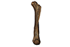
The present contribution contains the 3D virtual restoration of a Pliocene Lutrine right femur of Tobène, Senegal, described and figured in Lihoreau et al. (2021) : "A fossil terrestrial fauna from Tobène (Senegal) provides a unique early Pliocene window in Western Africa ". https://doi.org/10.1016/j.gr.2021.06.013
Indet indet SN-Tob-12-02 View specimen
|
M3#441Virtual restoration of SN-Tob-12-02 Type: "3D_surfaces"doi: 10.18563/m3.sf.441 state:published |
Download 3D surface file |
The present 3D Dataset contains the 3D models analyzed in the publication “Systematic and locomotor diversification of the Adapis group (Primates, Adapiformes) in the late Eocene of the Quercy (Southwest France), revealed by humeral remains”. In this paper, twenty humeral specimens from the old and new Quercy collections attributed to the fossil primates Adapis and Palaeolemur are described and analysed together. In this dataset only the scans of the fossils belonging to the collections of Université de Montpellier are provided.
In our paper (Marigó et al., 2019) we provide a qualitative and quantitative analysis of the different humeri, revealing that high variability is present within the “Adapis group” sample. Six different morphotypes are identified, confirming that what has often been called “Adapis parisiensis” is a mix of different species that present different locomotor adaptations.
Adapis sp. UM ROS 2-95 View specimen
|
M3#356Complete right humerus ROS 2-95 attributed to the Adapis group Type: "3D_surfaces"doi: 10.18563/m3.sf.356 state:published |
Download 3D surface file |
Adapis sp. UM ROS 2-536 View specimen
|
M3#357Proximal end of the right humerus ROS 2-536 attributed to the Adapis group Type: "3D_surfaces"doi: 10.18563/m3.sf.357 state:published |
Download 3D surface file |
Adapis sp. UM ROS 2-534 View specimen
|
M3#358Distal end of the left humerus ROS 2-534 attributed to the Adapis group Type: "3D_surfaces"doi: 10.18563/m3.sf.358 state:published |
Download 3D surface file |
Adapis sp. UM ROS 2-535 View specimen
|
M3#359Distal end of the left humerus ROS 2-535 attributed to the Adapis group Type: "3D_surfaces"doi: 10.18563/m3.sf.359 state:published |
Download 3D surface file |
Adapis sp. UM ROS 2-80 View specimen

|
M3#360Proximal end of the right humerus ROS 2-80 attributed to the Adapis group Type: "3D_surfaces"doi: 10.18563/m3.sf.360 state:published |
Download 3D surface file |
Adapis sp. UM ROS 2-79 View specimen
|
M3#361Distal end of the right humerus ROS 2-79 attributed to the Adapis group Type: "3D_surfaces"doi: 10.18563/m3.sf.361 state:published |
Download 3D surface file |
Adapis sp. UM ECA 1364 View specimen

|
M3#362Distal end of the left humerus ECA 1364 attributed to the Adapis group Type: "3D_surfaces"doi: 10.18563/m3.sf.362 state:published |
Download 3D surface file |
Adapis sp. UM ACQ-262 View specimen
|
M3#3733D model of ACQ 262. Humerus Type: "3D_surfaces"doi: 10.18563/m3.sf373 state:published |
Download 3D surface file |
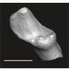
This note presents the 3D model of the hemi-mandible UM-PAT 159 of the MP7 Diacodexis species D. cf. gigasei and 3D models corresponding to the restoration of the ascending ramus, broken on the original specimen, and to a restoration of a complete mandible based on the preserved left hemi-mandible.
Diacodexis cf. gigasei UMPAT159 View specimen
|
M3#3153D models of UM PAT 159 after the restoration of the ascending ramus Type: "3D_surfaces"doi: 10.18563/m3.sf.315 state:published |
Download 3D surface file |
|
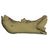
M3#316restoration of a complete mandible based on the preserved left hemi-mandible UM PAT 159 Type: "3D_surfaces"doi: 10.18563/m3.sf.316 state:published |
Download 3D surface file |
|
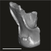
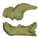
M3#3173D model of the hemi-mandible UM PAT 159 Type: "3D_surfaces"doi: 10.18563/m3.sf.317 state:published |
Download 3D surface file |
The present 3D Dataset contains the 3D models analyzed in: Hirose, A., Nakashima, T., Yamada, S., Uwabe, C., Kose, K., Takakuwa, T. 2012. Embryonic liver morphology and morphometry by magnetic resonance microscopic imaging. Anat Rec (Hoboken) 295, 51-59. doi: 10.1002/ar.21496
Homo sapiens KC-CS14LIV1387 View specimen
|
M3#64Human liver at Carnegie Stage (CS) 14 Type: "3D_surfaces"doi: 10.18563/m3.sf.64 state:published |
Download 3D surface file |
Homo sapiens KC-CS15LIV5074 View specimen
|
M3#65Human liver at Carnegie Stage (CS) 15 Type: "3D_surfaces"doi: 10.18563/m3.sf.65 state:published |
Download 3D surface file |
Homo sapiens KC-CS16LIV2578 View specimen
|
M3#66Human liver at Carnegie Stage (CS) 16 Type: "3D_surfaces"doi: 10.18563/m3.sf.66 state:published |
Download 3D surface file |
Homo sapiens KC-CS17LIV17832 View specimen
|
M3#67Human liver at Carnegie Stage (CS) 17 Type: "3D_surfaces"doi: 10.18563/m3.sf.67 state:published |
Download 3D surface file |
Homo sapiens KC-CS18LIV21124 View specimen
|
M3#68Human liver at Carnegie Stage (CS) 18 Type: "3D_surfaces"doi: 10.18563/m3.sf.68 state:published |
Download 3D surface file |
Homo sapiens KC-CS19LIV14353 View specimen

|
M3#69Human liver at Carnegie Stage (CS) 19 Type: "3D_surfaces"doi: 10.18563/m3.sf.69 state:published |
Download 3D surface file |
Homo sapiens KC-CS20LIV20701 View specimen
|
M3#70Human liver at Carnegie Stage (CS) 20 Type: "3D_surfaces"doi: 10.18563/m3.sf.70 state:published |
Download 3D surface file |
Homo sapiens KC-CS21LIV25858 View specimen
|
M3#71Human liver at Carnegie Stage (CS) 21 Type: "3D_surfaces"doi: 10.18563/m3.sf.71 state:published |
Download 3D surface file |
Homo sapiens KC-CS22LIV22226 View specimen
|
M3#72Human liver at Carnegie Stage (CS) 22 Type: "3D_surfaces"doi: 10.18563/m3.sf.72 state:published |
Download 3D surface file |
Homo sapiens KC-CS23LIV25704 View specimen
|
M3#73Human liver at Carnegie Stage (CS) 23 Type: "3D_surfaces"doi: 10.18563/m3.sf.73 state:published |
Download 3D surface file |
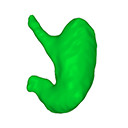
The present 3D Dataset contains the 3D models analyzed in: Kaigai N et al. Morphogenesis and three-dimensional movement of the stomach during the human embryonic period, Anat Rec (Hoboken). 2014 May;297(5):791-797. doi: 10.1002/ar.22833.
Homo sapiens KC-CS16STM27159 View specimen
|
M3#56computationally reconstructed stomach of the human embryo (M3#56_KC-CS16STM27159) at Carnegie Stage 16 (Crown Rump Length= 9.9mm). Type: "3D_surfaces"doi: 10.18563/m3.sf56 state:published |
Download 3D surface file |
Homo sapiens KC-CS17STM20383 View specimen
|
M3#57computationally reconstructed stomach of the human embryo (M3#57_KC-CS17STM20383) at Carnegie Stage 17 (Crown Rump Length= 12.3mm). Type: "3D_surfaces"doi: 10.18563/m3.sf57 state:published |
Download 3D surface file |
Homo sapiens KC-CS18STM21807 View specimen
|
M3#58computationally reconstructed stomach of the human embryo (M3#58_KC-CS18STM21807) at Carnegie Stage 18 (Crown Rump Length= 14.7mm). Type: "3D_surfaces"doi: 10.18563/m3.sf58 state:published |
Download 3D surface file |
Homo sapiens KC-CS19STM17998 View specimen
|
M3#59computationally reconstructed stomach of the human embryo (M3#59_KC-CS19STM17998) at Carnegie Stage 19 (Crown Rump Length was unmeasured ). Type: "3D_surfaces"doi: 10.18563/m3.sf59 state:published |
Download 3D surface file |
Homo sapiens KC-CS20STM20785 View specimen
|
M3#60computationally reconstructed stomach of the human embryo (M3#60_KC-CS20STM20785) at Carnegie Stage 20 (Crown Rump Length= 18.7 mm). Type: "3D_surfaces"doi: 10.18563/m3.sf60 state:published |
Download 3D surface file |
Homo sapiens KC-CS21STM24728 View specimen
|
M3#61computationally reconstructed stomach of the human embryo (M3#61_KC-CS21STM24728) at Carnegie Stage 21 (Crown Rump Length= 20.9 mm). Type: "3D_surfaces"doi: 10.18563/m3.sf61 state:published |
Download 3D surface file |
Homo sapiens KC-CS22STM26438 View specimen
|
M3#62computationally reconstructed stomach of the human embryo (M3#62_KC-CS22STM26438) at Carnegie Stage 22 (Crown Rump Length= 21.5 mm). Type: "3D_surfaces"doi: 10.18563/m3.sf62 state:published |
Download 3D surface file |
Homo sapiens KC-CS23STM20018 View specimen
|
M3#63computationally reconstructed stomach of the human embryo (M3#63_KC-CS23STM20018) at Carnegie Stage 23 (Crown Rump Length= 23.1 mm). Type: "3D_surfaces"doi: 10.18563/m3.sf63 state:published |
Download 3D surface file |
This contribution contains the 3D models described and figured in the following publication: Pujos F., Hautier L., Antoine P-O., Boivin M., Moison B, Salas-Gismondi R, Tejada J.V. , Varas-Malca R.M., Yans J., Marivaux L. (2025). Unexpected pampatheriid from the early Oligocene of Peruvian Amazonia: insights into the tropical differentiation of cingulate xenarthrans. Historical Biology.
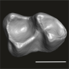
Bradypus tridactylus UM-ZOOL-V69 View specimen
|
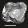
M3#1600Molariform and associated dentinal microstructure Type: "3D_surfaces"doi: 10.18563/m3.sf.1600 state:published |
Download 3D surface file |
Choloepus didactylus UM-ZOOL-V12 View specimen
|
M3#1601Molariform and associated dentinal microstructure Type: "3D_surfaces"doi: 10.18563/m3.sf.1601 state:published |
Download 3D surface file |
Dasypus mexicanus UM-ZOOL-2787 View specimen
|
M3#1602Molariform and associated dentinal microstructure Type: "3D_surfaces"doi: 10.18563/m3.sf.1602 state:published |
Download 3D surface file |
Tolypeutes matacus UM-ZOOL-2789 View specimen
|
M3#1603Molariform and associated dentinal microstructure Type: "3D_surfaces"doi: 10.18563/m3.sf.1603 state:published |
Download 3D surface file |
Euphractus sexcinctus UM-ZOOL-2790 View specimen
|
M3#1604Molariform and associated dentinal microstructure Type: "3D_surfaces"doi: 10.18563/m3.sf.1604 state:published |
Download 3D surface file |
Holmesina septrionalis UM-FLD-1 View specimen
|
M3#1605Molariform and associated dentinal microstructure Type: "3D_surfaces"doi: 10.18563/m3.sf.1605 state:published |
Download 3D surface file |
Megatherium sp. UM-TAR-1 View specimen
|
M3#1607Molariform and associated dentinal microstructure Type: "3D_surfaces"doi: 10.18563/m3.sf.1607 state:published |
Download 3D surface file |
Indet indet MUSM-3965 View specimen
|
M3#1606Molariform and associated dentinal microstructure Type: "3D_surfaces"doi: 10.18563/m3.sf.1606 state:published |
Download 3D surface file |
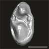
This contribution contains the 3D model described and figured in the following publication: Martin, T., Averianov, A. O., Schultz, J. A., & Schwermann, A. H. (2023). A stem therian mammal from the Lower Cretaceous of Germany. Journal of Vertebrate Paleontology, e2224848.
Spelaeomolitor speratus WMNM P99101 View specimen

|
M3#12573D_model_Spelaeomolitor_lower_molar Type: "3D_surfaces"doi: 10.18563/m3.sf.1257 state:published |
Download 3D surface file |
|
M3#1258CT imagestack (jpgs) and info data sheet (pca file) in one zip folder Type: "3D_CT"doi: 10.18563/m3.sf.1258 state:published |
Download CT data |
This contribution contains the three-dimensional digital models of the dental fossil material of anthropoid and strepsirrhine primates, discovered in Lower Oligocene detrital deposits outcropping in the Porto Rico and El Argoub areas, east of the Dakhla peninsula region (Atlantic Sahara; in the south of Morocco, near the northern border of Mauritania). These fossils were described, figured and discussed in the following publication: Marivaux et al. (2024), A new primate community from the earliest Oligocene of the Atlantic margin of Northwest Africa: Systematic, paleobiogeographic and paleoenvironmental implications. Journal of Human Evolution. https://doi.org/10.1016/j.jhevol.2024.103548
Catopithecus aff. browni DAK-Arg-087 View specimen
|
M3#1211Isolated right lower m3 (worn) Type: "3D_surfaces"doi: 10.18563/m3.sf.1211 state:published |
Download 3D surface file |
Catopithecus aff. browni DAK-Arg-088 View specimen
|
M3#1212Isolated right lower m2 (abraded/corroded) Type: "3D_surfaces"doi: 10.18563/m3.sf.1212 state:published |
Download 3D surface file |
Catopithecus aff. browni DAK-Arg-089 View specimen
|
M3#1213Isolated left lower m1 (worn) Type: "3D_surfaces"doi: 10.18563/m3.sf.1213 state:published |
Download 3D surface file |
Catopithecus aff. browni DAK-Pto-052 View specimen
|
M3#1214Isolated right lower m1 (pristine but lacking the mesiobuccal region) Type: "3D_surfaces"doi: 10.18563/m3.sf.1214 state:published |
Download 3D surface file |
Catopithecus aff. browni DAK-Arg-090 View specimen
|
M3#1215Isolated left upper P4 Type: "3D_surfaces"doi: 10.18563/m3.sf.1215 state:published |
Download 3D surface file |
Catopithecus aff. browni DAK-Arg-091 View specimen
|
M3#1216Isolated left upper M2 (worn and corroded) Type: "3D_surfaces"doi: 10.18563/m3.sf.1216 state:published |
Download 3D surface file |
Catopithecus aff. browni DAK-Pto-053 View specimen
|
M3#1217Isolated right upper M1 (lacking the buccal region) Type: "3D_surfaces"doi: 10.18563/m3.sf.1217 state:published |
Download 3D surface file |
Abuqatrania cf. basiodontos DAK-Arg-092 View specimen
|
M3#1218Isolated left lower c1 Type: "3D_surfaces"doi: 10.18563/m3.sf.1218 state:published |
Download 3D surface file |
?Propliopithecus sp. DAK-Pto-056 View specimen
|
M3#1219Isolated right lower m3 (fragment of talonid of a germ) Type: "3D_surfaces"doi: 10.18563/m3.sf.1219 state:published |
Download 3D surface file |
Abuqatrania cf. basiodontos DAK-Arg-093 View specimen
|
M3#1469Isolated right lower m1 Type: "3D_surfaces"doi: 10.18563/m3.sf.1469 state:published |
Download 3D surface file |
Abuqatrania cf. basiodontos DAK-Arg-094 View specimen
|
M3#1221Isolated left upper M1 or M2 (corroded, lacking the enamel cap [exposed dentine]) Type: "3D_surfaces"doi: 10.18563/m3.sf.1221 state:published |
Download 3D surface file |
Abuqatrania cf. basiodontos DAK-Arg-095 View specimen
|
M3#1222Isolated right lower i1 or i2 Type: "3D_surfaces"doi: 10.18563/m3.sf.1222 state:published |
Download 3D surface file |
Abuqatrania cf. basiodontos DAK-Arg-096 View specimen
|
M3#1223Isolated right lower p2 (worn apex) Type: "3D_surfaces"doi: 10.18563/m3.sf.1223 state:published |
Download 3D surface file |
Abuqatrania cf. basiodontos DAK-Arg-097 View specimen
|
M3#1224Isolated right lower p2 (worn apex and broken root) Type: "3D_surfaces"doi: 10.18563/m3.sf.1224 state:published |
Download 3D surface file |
Afrotarsius sp. DAK-Arg-098 View specimen
|
M3#1225Isolated left lower p3 Type: "3D_surfaces"doi: 10.18563/m3.sf.1225 state:published |
Download 3D surface file |
Afrotarsius sp. DAK-Pto-054 View specimen
|
M3#1226Isolated right lower m1 (abraded/corroded) Type: "3D_surfaces"doi: 10.18563/m3.sf.1226 state:published |
Download 3D surface file |
Orolemur mermozi DAK-Pto-055 View specimen
|
M3#1227Isolated right upper M1 or M2 (pristine, Holotype) Type: "3D_surfaces"doi: 10.18563/m3.sf.1227 state:published |
Download 3D surface file |
Wadilemur cf. elegans DAK-Arg-099 View specimen
|
M3#1228Isolated right lower m2 Type: "3D_surfaces"doi: 10.18563/m3.sf.1228 state:published |
Download 3D surface file |
cf. 'Anchomomys' milleri DAK-Arg-100 View specimen
|
M3#1229Isolated right lower c1 Type: "3D_surfaces"doi: 10.18563/m3.sf.1229 state:published |
Download 3D surface file |
Abuqatrania cf. basiodontos DAK-Arg-101 View specimen
|
M3#1396Isolated left upper M3 (abraded) Type: "3D_surfaces"doi: 10.18563/m3.sf.1396 state:published |
Download 3D surface file |
Orogalago saintexuperyi DAK-Arg-102 View specimen
|
M3#1397Isolated left lower m2 Type: "3D_surfaces"doi: 10.18563/m3.sf.1397 state:published |
Download 3D surface file |
Wadilemur cf. elegans DAK-Arg-103 View specimen
|
M3#1473Isolated right upper M1 or M2 (lacking the mesial and buccal regions) Type: "3D_surfaces"doi: 10.18563/m3.sf.1473 state:published |
Download 3D surface file |
This contribution contains the 3D models described and figured in the following publication: Bonis et al. 2023. A new large pantherine and a sabre-toothed cat (Mammalia, Carnivora, Felidae) from the late Miocene hominoid-bearing Khorat sand pits, Nakhon Ratchasima Province, northeastern Thailand. The Science of Nature 110(5):42. https://doi.org/10.1007/s00114-023-01867-4
Pachypanthera piriyai CUF-KR-1 View specimen
|
M3#1209Holotype of Pachypanthera piriyai, a left hemi-mandible with alveoli for i1-i3 and canine, roots of p3, p4 and partially broken off m1 crown. Type: "3D_surfaces"doi: 10.18563/m3.sf.1209 state:published |
Download 3D surface file |
Pachypanthera piriyai CUF-KR-2 View specimen
|
M3#1210Paratype of Pachypanthera piriyai, a right hemi-maxilla with P3-P4, alveoli of C and M1, root of P2 Type: "3D_surfaces"doi: 10.18563/m3.sf.1210 state:published |
Download 3D surface file |
This contribution contains 3D models of the holotype of a new species of long-nosed armadillos, the Guianan long-nosed armadillo (Dasypus guianensis) described in the following publication: Barthe M., Rancilhac L., Arteaga M. C., Feijó A., Tilak M.-K., Justy F., Loughry W. J., McDonough C. M., de Thoisy B., Catzeflis F., Billet G., Hautier L., Nabholz B., and Delsuc F. 2024. Exon capture museomics deciphers the nine-banded armadillo species complex and identifies a new species endemic to the Guiana Shield. Systematic Biology, syae027. https://doi.org/10.1093/sysbio/syae027
Dasypus guianensis MNHN-ZM-MO-2001-1317 View specimen
|
M3#1200Skeleton and carapace Type: "3D_surfaces"doi: 10.18563/m3.sf.1200 state:published |
Download 3D surface file |
|
M3#1201Frontal sinuses Type: "3D_surfaces"doi: 10.18563/m3.sf.1201 state:published |
Download 3D surface file |
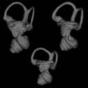
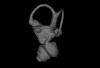
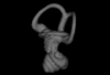
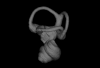
This contribution contains the three-dimensional models of the inner ear of the hetaxodontid rodents Amblyrhiza, Clidomys and Elasmodontomys from the West Indies. These specimens were analyzed and discussed in : The inner ear of caviomorph rodents: phylogenetic implications and application to extinct West Indian taxa.
Amblyrhiza inundata 11842 View specimen
|
M3#11543D surface of the left-oriented inner ear of Amblyrhiza. Type: "3D_surfaces"doi: 10.18563/m3.sf.1154 state:published |
Download 3D surface file |
Clidomys sp NA View specimen
|
M3#11553D surface of the left-oriented inner ear of Clidomys sp. Type: "3D_surfaces"doi: 10.18563/m3.sf.1155 state:published |
Download 3D surface file |
Elasmodontomys obliquus 17127 View specimen
|
M3#11563D surface of the left-oriented inner ear of Elasmodontomys obliquus. Type: "3D_surfaces"doi: 10.18563/m3.sf.1156 state:published |
Download 3D surface file |
The present 3D Dataset contains the 3D model analyzed in the following publication: Carolina A. Hoffmann, A. G. Martinelli & M. B. Andrade. 2023. Anatomy of the holotype of “Probelesodon” kitchingi revisited, a chiniquodontid cynodont (Synapsida, Probainognathia) from the early Late Triassic of southern Brazil, Journal of Paleontology
Probelesodon kitchingi MCP 1600 PV View specimen
|
M3#11513D models of the skull with segmented bones and without the segmentation. colormap and orientation files also added. Type: "3D_surfaces"doi: 10.18563/m3.sf.1151 state:published |
Download 3D surface file |

This contribution contains the 3D models analyzed in Müller et al. (2021) “Pushing the boundary? Testing the ‘functional elongation hypothesis’ of the giraffe’s neck”.
Aepyceros melampus ZFMK 2001.278 View specimen

|
M3#643Vertebrae C7, T1 Type: "3D_surfaces"doi: 10.18563/m3.sf.643 state:published |
Download 3D surface file |
Giraffa camelopardalis ZMB 66393 View specimen

|
M3#644Vertebrae Type: "3D_surfaces"doi: 10.18563/m3.sf.644 state:published |
Download 3D surface file |
Giraffa camelopardalis ZSM 1967/17 View specimen

|
M3#645Vertebrae Type: "3D_surfaces"doi: 10.18563/m3.sf.645 state:published |
Download 3D surface file |
Giraffa camelopardalis ZSM 1981/19 View specimen

|
M3#646C3, C4, C5, C6, C7, T1, T2 Type: "3D_surfaces"doi: 10.18563/m3.sf.646 state:published |
Download 3D surface file |
Giraffa camelopardalis KMDA M-10861 View specimen

|
M3#647C3, C4, C5, C6, C7, T1, T2. Acquired via laser scanner. Type: "3D_surfaces"doi: 10.18563/m3.sf.647 state:published |
Download 3D surface file |
Giraffa camelopardalis SMF 84214 View specimen

|
M3#648C7, T1. Warning : photogrammetric models (unit scale is CM, not MM). Type: "3D_surfaces"doi: 10.18563/m3.sf.648 state:published |
Download 3D surface file |
Giraffa camelopardalis SMF 78299 View specimen

|
M3#649C7, T1. Warning : unscaled photogrammetric 3D models (unknown size). Type: "3D_surfaces"doi: 10.18563/m3.sf.649 state:published |
Download 3D surface file |
Giraffa camelopardalis SMF o. N View specimen

|
M3#650C7, T1. Warning : unscaled photogrammetric 3D models (unknown size). Type: "3D_surfaces"doi: 10.18563/m3.sf.650 state:published |
Download 3D surface file |
Giraffa camelopardalis SMNS 19138 View specimen
|
M3#671C7, T1. Warning : unscaled photogrammetric 3D models (unknown size). Type: "3D_surfaces"doi: 10.18563/m3.sf.671 state:published |
Download 3D surface file |
Okapia johnstoni ZMB 62086 View specimen

|
M3#651C3, C4, C5, C6, C7, T1, T2 Type: "3D_surfaces"doi: 10.18563/m3.sf.651 state:published |
Download 3D surface file |
Okapia johnstoni ZMB 70325 View specimen

|
M3#652C3, C4, C5, C6, C7, T1, T2 Type: "3D_surfaces"doi: 10.18563/m3.sf.652 state:published |
Download 3D surface file |
Sivatherium giganteum NHMUK 15707 View specimen
|
M3#653C7. Warning : unscaled photogrammetric 3D model (unknown size). Type: "3D_surfaces"doi: 10.18563/m3.sf.653 state:published |
Download 3D surface file |
Sivatherium giganteum NHMUK 15297 View specimen

|
M3#654T1. Warning : unscaled photogrammetric 3D model (unknown size). Type: "3D_surfaces"doi: 10.18563/m3.sf.654 state:published |
Download 3D surface file |
Cervus elaphus ZMB 47502 View specimen

|
M3#655C3, C4, C5, C6, C7, T1, T2 Type: "3D_surfaces"doi: 10.18563/m3.sf.655 state:published |
Download 3D surface file |
Axis axis SMF 1450 View specimen

|
M3#656C7, T1 Type: "3D_surfaces"doi: 10.18563/m3.sf.656 state:published |
Download 3D surface file |
Cervus nippon SMF 4368 View specimen
|
M3#657C7, T1 Type: "3D_surfaces"doi: 10.18563/m3.sf.657 state:published |
Download 3D surface file |
Capreolus capreolus SMF 79852 View specimen

|
M3#658C7, T1 Type: "3D_surfaces"doi: 10.18563/m3.sf.658 state:published |
Download 3D surface file |
Capreolus capreolus ZFMK 67.237 View specimen

|
M3#659C7, T1 Type: "3D_surfaces"doi: 10.18563/m3.sf.659 state:published |
Download 3D surface file |
Muntiacus reevesi SMF 92954 View specimen

|
M3#660C7, T1 Type: "3D_surfaces"doi: 10.18563/m3.sf.660 state:published |
Download 3D surface file |
Muntiacus reevesi SMF 92332 View specimen

|
M3#661C7, T1 Type: "3D_surfaces"doi: 10.18563/m3.sf.661 state:published |
Download 3D surface file |
Alces alces SMF 35549 View specimen

|
M3#662C7, T1 Type: "3D_surfaces"doi: 10.18563/m3.sf.662 state:published |
Download 3D surface file |
Dama dama ZFMK 86.125 View specimen

|
M3#663C7, T1 Type: "3D_surfaces"doi: 10.18563/m3.sf.663 state:published |
Download 3D surface file |
Antilope cervicapra ZMB 78829 View specimen

|
M3#664C3, C4, C5, C6, C7, T1, T2 Type: "3D_surfaces"doi: 10.18563/m3.sf.664 state:published |
Download 3D surface file |
Bison bonasus SMNS 2998 View specimen

|
M3#665C7, T1. Warning : unscaled photogrammetric 3D models (unknown size). Type: "3D_surfaces"doi: 10.18563/m3.sf.665 state:published |
Download 3D surface file |
Nanger dama SMF 74435 View specimen

|
M3#666C7, T1 Type: "3D_surfaces"doi: 10.18563/m3.sf.666 state:published |
Download 3D surface file |
Litocranius walleri SMF 23747 View specimen

|
M3#667C7, T1 Type: "3D_surfaces"doi: 10.18563/m3.sf.667 state:published |
Download 3D surface file |
Litocranius walleri SMF 23749 View specimen

|
M3#669C7, T1 Type: "3D_surfaces"doi: 10.18563/m3.sf.669 state:published |
Download 3D surface file |
Tragelaphus eurycerus SMF 95875 View specimen

|
M3#670C7, T1 Type: "3D_surfaces"doi: 10.18563/m3.sf.670 state:published |
Download 3D surface file |
Bos javanicus SMF 64934 View specimen

|
M3#672C7, T1 Type: "3D_surfaces"doi: 10.18563/m3.sf.672 state:published |
Download 3D surface file |
Ovis aries ZFMK 1982.338 View specimen

|
M3#673C7, T1 Type: "3D_surfaces"doi: 10.18563/m3.sf.673 state:published |
Download 3D surface file |
Rupicapra rupicapra ZFMK 72.367 View specimen

|
M3#674C7, T1 Type: "3D_surfaces"doi: 10.18563/m3.sf.674 state:published |
Download 3D surface file |
Kobus ellipsiprymnus SMNS 4443 View specimen

|
M3#675C7, T1 Type: "3D_surfaces"doi: 10.18563/m3.sf.675 state:published |
Download 3D surface file |
Sylvicapra grimmia SMNS 15292 View specimen

|
M3#676C7, T1 Type: "3D_surfaces"doi: 10.18563/m3.sf.676 state:published |
Download 3D surface file |
Syncerus caffer SMNS 7347 View specimen

|
M3#677C7, T1. Warning : unscaled photogrammetric 3D models (unknown size). Type: "3D_surfaces"doi: 10.18563/m3.sf.677 state:published |
Download 3D surface file |
Procapra gutturosa SMNS 5796 View specimen

|
M3#678C7, T1 Type: "3D_surfaces"doi: 10.18563/m3.sf.678 state:published |
Download 3D surface file |
Damaliscus pygargus SMNS 21617 View specimen

|
M3#679C7, T1 Type: "3D_surfaces"doi: 10.18563/m3.sf.679 state:published |
Download 3D surface file |
Madoqua kirkii SMNS 4432 View specimen

|
M3#680C7, T1 Type: "3D_surfaces"doi: 10.18563/m3.sf.680 state:published |
Download 3D surface file |
Bubalus mindorensis SMNS 2054 View specimen

|
M3#681C7, T1. Warning : unscaled photogrammetric 3D models (unknown size). Type: "3D_surfaces"doi: 10.18563/m3.sf.681 state:published |
Download 3D surface file |
Capra hircus SMNS 51328 View specimen

|
M3#682C7, T1 Type: "3D_surfaces"doi: 10.18563/m3.sf.682 state:published |
Download 3D surface file |
Connochaetes taurinus SMNS 4442 View specimen

|
M3#683C7, T1. Warning : unscaled photogrammetric 3D models (unknown size). Type: "3D_surfaces"doi: 10.18563/m3.sf.683 state:published |
Download 3D surface file |
Antilocapra americana ZSM 1964/218 View specimen

|
M3#684C3, C4, C5, C6, C7, T1, T2 Type: "3D_surfaces"doi: 10.18563/m3.sf.684 state:published |
Download 3D surface file |
Antilocapra americana ZMB 77281 View specimen

|
M3#685C7, T1 Type: "3D_surfaces"doi: 10.18563/m3.sf.685 state:published |
Download 3D surface file |
Moschus moschiferus ZMB 62080 View specimen

|
M3#686C3, C4, C5, C6, C7, T1, T2 Type: "3D_surfaces"doi: 10.18563/m3.sf.686 state:published |
Download 3D surface file |
Moschus moschiferus ZMB 60367 View specimen

|
M3#687C7, T1 Type: "3D_surfaces"doi: 10.18563/m3.sf.687 state:published |
Download 3D surface file |
Moschus moschiferus ZMB 51830 View specimen

|
M3#688C7, T1 Type: "3D_surfaces"doi: 10.18563/m3.sf.688 state:published |
Download 3D surface file |
Tragulus javanicus SMF 82179 View specimen

|
M3#689C7, T1 Type: "3D_surfaces"doi: 10.18563/m3.sf.689 state:published |
Download 3D surface file |
Tragulus javanicus ZMB 86222 View specimen

|
M3#690C7, T1 Type: "3D_surfaces"doi: 10.18563/m3.sf.690 state:published |
Download 3D surface file |
Tragulus sp. ZMB o. N. View specimen

|
M3#691C7, T1 Type: "3D_surfaces"doi: 10.18563/m3.sf.691 state:published |
Download 3D surface file |
Hyemoschus aquaticus ZMB 71071 View specimen

|
M3#692C7, T1 Type: "3D_surfaces"doi: 10.18563/m3.sf.692 state:published |
Download 3D surface file |
Hyemoschus aquaticus ZMB 103235 View specimen

|
M3#693C7, T1 Type: "3D_surfaces"doi: 10.18563/m3.sf.693 state:published |
Download 3D surface file |
Vicugna vicugna SMF 94752 View specimen

|
M3#694C7, T1 Type: "3D_surfaces"doi: 10.18563/m3.sf.694 state:published |
Download 3D surface file |
Camelus dromedarius SMF 70473 View specimen

|
M3#695C7, T1. Warning : unscaled photogrammetric 3D models (unknown size). Type: "3D_surfaces"doi: 10.18563/m3.sf.695 state:published |
Download 3D surface file |
Camelus bactrianus SMF 25542 View specimen

|
M3#696C7, T1. Warning : unscaled photogrammetric 3D models (unknown size). Type: "3D_surfaces"doi: 10.18563/m3.sf.696 state:published |
Download 3D surface file |
Lama glama SMNS 31175 View specimen

|
M3#697C7, T1 Type: "3D_surfaces"doi: 10.18563/m3.sf.697 state:published |
Download 3D surface file |
Vicugna pacos SMNS 46255 View specimen

|
M3#698C7, T1 Type: "3D_surfaces"doi: 10.18563/m3.sf.698 state:published |
Download 3D surface file |
Vicugna pacos SMNS 7349 View specimen

|
M3#699C7, T1 Type: "3D_surfaces"doi: 10.18563/m3.sf.699 state:published |
Download 3D surface file |
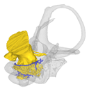
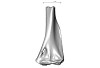
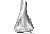
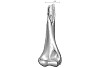
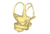
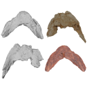
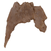
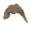
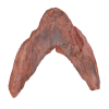
This contribution contains the 3D models described and figured in the publication entitled "The petrosal and bony labyrinth of Diplobune minor, an enigmatic Artiodactyla from the Oligocene of Western Europe" by Orliac, Araújo, and Lihoreau published in Journal of Morphology (Orliac et al. 2017) https://doi.org/10.1002/jmor.20702.
Diplobune minor UM ITD 1079 View specimen
|
M3#138right bony labyrinth of Diplobune minor from Itardies, France Type: "3D_surfaces"doi: 10.18563/m3.sf.138 state:published |
Download 3D surface file |
|
M3#139right isolated petrosal of Diplobune minor from Itardies, France Type: "3D_surfaces"doi: 10.18563/m3.sf.139 state:published |
Download 3D surface file |
Diplobune minor UM ITD 1080 View specimen
|
M3#140left bony labyrinth of Diplobune minor from Itardies, France Type: "3D_surfaces"doi: 10.18563/m3.sf.140 state:published |
Download 3D surface file |
|
M3#141left isolated petrosal of Diplobune minor from Itardies, France Type: "3D_surfaces"doi: 10.18563/m3.sf.141 state:published |
Download 3D surface file |
Diplobune minor UM ITD 1081 View specimen
|
M3#142right bony labyrinth and associated nerves and veins of Diplobune minor from Itardies, France Type: "3D_surfaces"doi: 10.18563/m3.sf.142 state:published |
Download 3D surface file |
|
M3#143right isolated petrosal of Diplobune minor from Itardies, France Type: "3D_surfaces"doi: 10.18563/m3.sf.143 state:published |
Download 3D surface file |
Diplobune minor UM ITD 1083 View specimen
|
M3#144left bony labyrinth of Diplobune minor from Itardies, France Type: "3D_surfaces"doi: 10.18563/m3.sf.144 state:published |
Download 3D surface file |
|
M3#145left petrosal of Diplobune minor from Itardies, France Type: "3D_surfaces"doi: 10.18563/m3.sf.145 state:published |
Download 3D surface file |
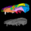
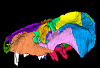
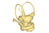
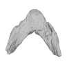
This contribution contains the 3D model described and figured in the following publication: Hautier L, Sarr R, Lihoreau F, Tabuce R, Marwan Hameh P. 2014. First record of the family Protocetidae in the Lutetian of Senegal (West Africa). Palaeovertebrata 38(2)-e2
indet. indet. SN103 View specimen
|
M3#5SN103, partial left innominate. Age and occurrence – Taïba Formation, Lutetian of the near Taïba Ndiaye, quarry of the Industries Chimiques du Sénégal (ICS) Type: "3D_surfaces"doi: 10.18563/m3.sf5 state:published |
Download 3D surface file |
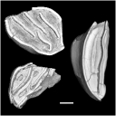
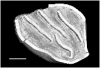
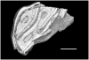
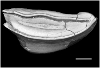
The 3D dataset presented in this article provides the 3D models of two Chelonioidea turtles dentaries from the Paleocene of France described in: Lapparent de Broin F. de, Marek H., Barrier P. & Gagnaison C. 2025. — Euclastidae n. fam. (Chelonioidea) et première mention d’Euclastes Cope, 1867 dans le Paléocène du bassin de Paris (France). Geodiversitas 47 (10): 409-464. https://doi.org/10.5252/geodiversitas2025v47a10.
Euclastes wielandi ULB-04A21-10 View specimen
|
M3#1791Euclastes wielandi Type: "3D_surfaces"doi: 10.18563/m3.sf.1791 state:published |
Download 3D surface file |
Euclastes wielandi MNHN.F.BPT52 View specimen
|
M3#1709Euclastes wielandi (cast) Type: "3D_surfaces"doi: 10.18563/m3.sf.1709 state:published |
Download 3D surface file |
Euclastes wielandi ULB-04A21-11 View specimen
|
M3#1792Euclastes montenati nov. sp. Type: "3D_surfaces"doi: 10.18563/m3.sf.1792 state:published |
Download 3D surface file |
Euclastes wielandi MNHN.F.BPT53 View specimen
|
M3#1711Euclastes montenati (cast) Type: "3D_surfaces"doi: 10.18563/m3.sf.1711 state:published |
Download 3D surface file |
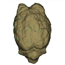
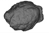
The present 3D Dataset contains the 3D model of the endocranial cast of Palaeolama sp. from the mid-Pleistocene (~1.2 Mya) of South America, analyzed in Balcarcel et al. 2023.
Palaeolama sp. PIMUZ A/V 4091 View specimen
|
M3#11283D model of a natural endocast Type: "3D_surfaces"doi: 10.18563/m3.sf.1128 state:published |
Download 3D surface file |
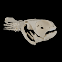
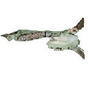
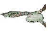
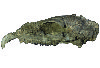
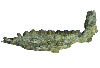
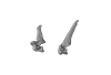
The present 3D Dataset contains 3D models of the cranial, visceral, and pectoral endoskeleton of Iniopera, an iniopterygian stem-group holocephalan from the Pennsylvanian of the USA. These data formed the basis for the analyses carried out in Dearden et al. (2023) “Evidence for high-performance suction feeding in the Pennsylvanian stem-group holocephalan Iniopera” PNAS.
Iniopera sp. KUNHM 22060, 158289 View specimen
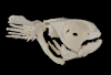
|
M3#1034plys of the head endoskeleton of Iniopera sp. Type: "3D_surfaces"doi: 10.18563/m3.sf.1034 state:published |
Download 3D surface file |


