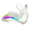
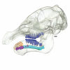
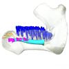
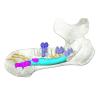
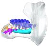
Explodable 3D Dog Skull for Veterinary Education
3D models of Ocnotherium skull
3D models of Kalakocetus, the earliest Cetacea
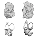
3D GM dataset of bird skeletal variation
Skeletal embryonic development in the catshark
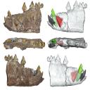
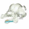
Bony connexions of the petrosal bone of extant hippos
bony labyrinth (14) , inner ear (11) , geometric morphometrics (10) , CT-scan (10) , Eocene (10) , Micro-CT (9) , Miocene (8)
Lionel Hautier (24) , Maëva Judith Orliac (23) , Laurent Marivaux (18) , Renaud Lebrun (14) , Rodolphe Tabuce (14) , Pierre-Olivier Antoine (13) , Bastien Mennecart (13)
|
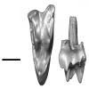
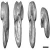
3D models related to the publication: Morphological study of the anterior dentition in Raoellidae (Mammalia, Artiodactyla), new insight on their dietary habits
|
|
M3#1505Right i1 Type: "3D_surfaces"doi: 10.18563/m3.sf.1505 state:published |
Download 3D surface file |
Indohyus indirae GU/RJ/32 View specimen
|
M3#1506Right i1 Type: "3D_surfaces"doi: 10.18563/m3.sf.1506 state:published |
Download 3D surface file |
Indohyus indirae GU/RJ/16 View specimen
|
M3#1507Left I3 Type: "3D_surfaces"doi: 10.18563/m3.sf.1507 state:published |
Download 3D surface file |
Indohyus indirae GU/RJ/23 View specimen
|
M3#1508Left I2 Type: "3D_surfaces"doi: 10.18563/m3.sf.1508 state:published |
Download 3D surface file |
Indohyus indirae GU/RJ/25 View specimen
|
M3#1509Left I1 Type: "3D_surfaces"doi: 10.18563/m3.sf.1509 state:published |
Download 3D surface file |
Indohyus indirae GU/RJ/26 View specimen
|
M3#1510Right I3 Type: "3D_surfaces"doi: 10.18563/m3.sf.1510 state:published |
Download 3D surface file |
Indohyus indirae GU/RJ/57 View specimen
|
M3#1511Left I1 Type: "3D_surfaces"doi: 10.18563/m3.sf.1511 state:published |
Download 3D surface file |
Indohyus indirae GU/RJ/61 View specimen
|
M3#1512Right upper canine Type: "3D_surfaces"doi: 10.18563/m3.sf.1512 state:published |
Download 3D surface file |
Indohyus indirae GU/RJ/63 View specimen
|
M3#1513Left upper canine Type: "3D_surfaces"doi: 10.18563/m3.sf.1513 state:published |
Download 3D surface file |
Indohyus indirae GU/RJ/74 View specimen
|
M3#1514Left upper canine Type: "3D_surfaces"doi: 10.18563/m3.sf.1514 state:published |
Download 3D surface file |
Indohyus indirae GU/RJ/439 View specimen
|
M3#1515Left upper canine Type: "3D_surfaces"doi: 10.18563/m3.sf.1515 state:published |
Download 3D surface file |
Indohyus indirae GU/RJ/457 View specimen
|
M3#1516Left upper canine Type: "3D_surfaces"doi: 10.18563/m3.sf.1516 state:published |
Download 3D surface file |
Indohyus indirae GU/RJ/846 View specimen
|
M3#1517Left upper canine Type: "3D_surfaces"doi: 10.18563/m3.sf.1517 state:published |
Download 3D surface file |
Indohyus indirae GU/RJ/822 View specimen
|
M3#1518Right fragmentary maxillary with decidual canine and I3 Type: "3D_surfaces"doi: 10.18563/m3.sf.1518 state:published |
Download 3D surface file |
Indohyus indirae GU/RJ/824 View specimen
|
M3#1519Right fragmentary mandible with lower canine and small part of the i3 Type: "3D_surfaces"doi: 10.18563/m3.sf.1519 state:published |
Download 3D surface file |
Indohyus indirae GU/RJ/838 View specimen
|
M3#1520Right fragmentary mandible with permanent i2, i3 and canine and small part of the root of the decidual i3 Type: "3D_surfaces"doi: 10.18563/m3.sf.1520 state:published |
Download 3D surface file |
Indohyus indirae GU/RJ/842 View specimen
|
M3#1521Left fragmentary mandible with decidual and permanent canine Type: "3D_surfaces"doi: 10.18563/m3.sf.1521 state:published |
Download 3D surface file |
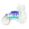
Indohyus indirae GU/RJ/26,32,56,57,457,822,838,842 : Composite Anterior dentition View specimen
|
M3#15293D composite reconstruction of the anterior dentition of Indohyus indirae with GU/RJ/57 (I1), 56 (I2), 26 (I3), 457 (Upper canine), 822 (P1), 32 (i1), 838 (i2, i3 and lower canine) and 842 (p1) Type: "3D_surfaces"doi: 10.18563/m3.sf.1529 state:published |
Download 3D surface file |
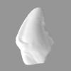
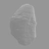
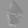
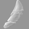
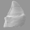
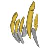
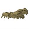
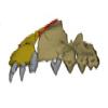
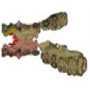
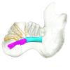
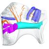
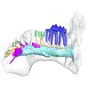
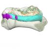
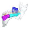
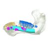
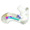
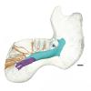
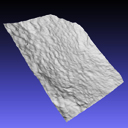
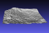
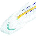
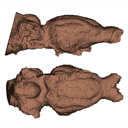
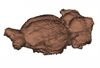
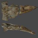
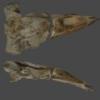
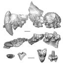
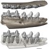
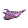
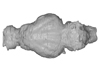
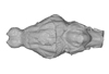
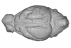
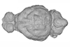
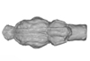
In this work, we digitally restore the snout of the raoellide Khirtharia inflata from the Kalakot area (Rajouri District, Jammu & Kashmir, India). Raoellids are small, semiaquatic ungulates closely related to cetaceans. The specimen is fairly complete and preserves left and right maxillaries, left premaxillary, and part of the anterior and jugal dentition. The digital restoration of this quite complete but deformed specimen of Khirtharia inflata is a welcome addition to the data available for raoellids and will be used to further the understanding of the origins of cetaceans.
Khirtharia inflata GU/RJ/157 View specimen
|
M3#1454deformed partial skull Type: "3D_surfaces"doi: 10.18563/m3.sf.1454 state:published |
Download 3D surface file |
|
M3#1455reconstruction of half snout Type: "3D_surfaces"doi: 10.18563/m3.sf.1455 state:published |
Download 3D surface file |
|
M3#1456reconstruction of complete snout Type: "3D_surfaces"doi: 10.18563/m3.sf.1456 state:published |
Download 3D surface file |
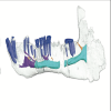
This contribution contains the 3D models described and figured in the following publication: Hautier L, Gomes Rodrigues H, Ferreira-Cardoso S, Emerling CA, Porcher M-L, Asher R, Portela Miguez R, Delsuc F. 2023. From teeth to pad: tooth loss and development of keratinous structures in sirenians. Proceedings of the Royal Society B. https://doi.org/10.1098/rspb.2023.1932
Dugong dugon 2005.51 View specimen

|
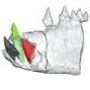
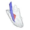
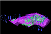
M3#1275Internal mandibular morphology. Orange = dorsal canaliculi; purple = mental branches; cyan = mandibular canal; dark blue = teeth; green = tooth alveoli. Type: "3D_surfaces"doi: 10.18563/m3.sf.1275 state:published |
Download 3D surface file |
Dugong dugon 2023.66 View specimen
|
M3#1274Internal mandibular morphology. Orange = dorsal canaliculi; purple = mental branches; cyan = mandibular canal; dark blue = teeth; green = tooth alveoli. Type: "3D_surfaces"doi: 10.18563/m3.sf.1274 state:published |
Download 3D surface file |
Dugong dugon 5386 View specimen
|
M3#1276Internal mandibular morphology. Orange = dorsal canaliculi; purple = mental branches; cyan = mandibular canal; dark blue = teeth; green = tooth alveoli. Type: "3D_surfaces"doi: 10.18563/m3.sf.1276 state:published |
Download 3D surface file |
Dugong dugon 1848.8.29.7/GERM 1027g View specimen
|
M3#1277Internal mandibular morphology. Orange = dorsal canaliculi; purple = mental branches; cyan = mandibular canal; dark blue = teeth. Type: "3D_surfaces"doi: 10.18563/m3.sf.1277 state:published |
Download 3D surface file |
Dugong dugon 1991.413 View specimen
|
M3#1278Internal mandibular morphology. Orange = dorsal canaliculi; purple = mental branches; cyan = mandibular canal; dark blue = teeth. Type: "3D_surfaces"doi: 10.18563/m3.sf.1278 state:published |
Download 3D surface file |
Dugong dugon 1991.427 View specimen
|
M3#1279Internal mandibular morphology. Orange = dorsal canaliculi; purple = mental branches; cyan = mandibular canal; dark blue = teeth. Type: "3D_surfaces"doi: 10.18563/m3.sf.1279 state:published |
Download 3D surface file |
Dugong dugon 2017-3-9 View specimen
|
M3#1280Internal mandibular morphology. Orange = dorsal canaliculi; purple = mental branches; cyan = mandibular canal; dark blue = teeth. Type: "3D_surfaces"doi: 10.18563/m3.sf.1280 state:published |
Download 3D surface file |
Eosiren lybica 1913-22 View specimen
|
M3#1281Internal mandibular morphology. Orange = dorsal canaliculi; purple = mental branches; cyan = mandibular canal; dark blue = teeth; green = tooth alveoli. Type: "3D_surfaces"doi: 10.18563/m3.sf.1281 state:published |
Download 3D surface file |
Halitherium taulannense RGHP C001 View specimen
|
M3#1282Internal mandibular morphology. Cyan = mandibular canal; dark blue = teeth. Type: "3D_surfaces"doi: 10.18563/m3.sf.1282 state:published |
Download 3D surface file |
Halitherium taulannense RGHP C009 View specimen
|
M3#1283Internal mandibular morphology. Orange = dorsal canaliculi; purple = mental branches; cyan = mandibular canal; dark blue = teeth; green = tooth alveoli. Type: "3D_surfaces"doi: 10.18563/m3.sf.1283 state:published |
Download 3D surface file |
Hydrodamalis gigas 1947.10.21.1 View specimen
|
M3#1284Internal mandibular morphology. Orange = dorsal canaliculi; purple = mental branches; cyan = mandibular canal Type: "3D_surfaces"doi: 10.18563/m3.sf.1284 state:published |
Download 3D surface file |
Hydrodamalis gigas C1021 View specimen
|
M3#1285Anterior part of the mandible Type: "3D_surfaces"doi: 10.18563/m3.sf.1285 state:published |
Download 3D surface file |

|
M3#1286Posterior part of the mandible Type: "3D_surfaces"doi: 10.18563/m3.sf.1286 state:published |
Download 3D surface file |
Hydrodamalis gigas 2023.67 View specimen
|
M3#1287Internal mandibular morphology. Orange = dorsal canaliculi; purple = mental branches; cyan = mandibular canal Type: "3D_surfaces"doi: 10.18563/m3.sf.1287 state:published |
Download 3D surface file |
Libysiren sickenbergi M.82429 View specimen
|
M3#1288Internal mandibular morphology. Orange = dorsal canaliculi; purple = mental branches; cyan = mandibular canal; dark blue = teeth; green = tooth alveoli. Type: "3D_surfaces"doi: 10.18563/m3.sf.1288 state:published |
Download 3D surface file |
Libysiren sickenbergi M.45675 View specimen
|
M3#1289Internal mandibular morphology. Orange = dorsal canaliculi; purple = mental branches; cyan = mandibular canal; dark blue = teeth; green = tooth alveoli. Type: "3D_surfaces"doi: 10.18563/m3.sf.1289 state:published |
Download 3D surface file |
Prorastomus sirenoides OR.448976 View specimen
|
M3#1290Internal morphology of the left mandible. Orange = dorsal canaliculi; purple = mental branches; cyan = mandibular canal; dark blue = teeth; green = tooth alveoli. Type: "3D_surfaces"doi: 10.18563/m3.sf.1290 state:published |
Download 3D surface file |
|
M3#1304Internal morphology of the right mandible. Orange = dorsal canaliculi; purple = mental branches; cyan = mandibular canal; dark blue = teeth. Type: "3D_surfaces"doi: 10.18563/m3.sf.1304 state:published |
Download 3D surface file |
Ribodon limbatus M.7073 View specimen
|
M3#1292Internal mandibular morphology. Orange = dorsal canaliculi; purple = mental branches; cyan = mandibular canal; dark blue = teeth; green = tooth alveoli. Type: "3D_surfaces"doi: 10.18563/m3.sf.1292 state:published |
Download 3D surface file |
Rytiodus capgrandi PAL2017-8-1 View specimen
|
M3#1293Internal mandibular morphology. Orange = dorsal canaliculi; purple = mental branches; cyan = mandibular canal; dark blue = teeth; green = tooth alveoli. Type: "3D_surfaces"doi: 10.18563/m3.sf.1293 state:published |
Download 3D surface file |
Trichechus inunguis 1868.12.19.2 View specimen
|
M3#1294Internal mandibular morphology. Orange = dorsal canaliculi; purple = mental branches; cyan = mandibular canal; dark blue = teeth; green = tooth alveoli. Type: "3D_surfaces"doi: 10.18563/m3.sf.1294 state:published |
Download 3D surface file |
Trichechus manatus 1843.3.10.12 View specimen
|
M3#1295Internal mandibular morphology. Orange = dorsal canaliculi; purple = mental branches; cyan = mandibular canal; dark blue = teeth. Type: "3D_surfaces"doi: 10.18563/m3.sf.1295 state:published |
Download 3D surface file |
Trichechus manatus 1864.6.5.1 View specimen
|
M3#1296Internal mandibular morphology. Orange = dorsal canaliculi; purple = mental branches; cyan = mandibular canal; dark blue = teeth; green = tooth alveoli. Type: "3D_surfaces"doi: 10.18563/m3.sf.1296 state:published |
Download 3D surface file |
Trichechus manatus 1950.1.23.1 View specimen
|
M3#1297Internal mandibular morphology. Orange = dorsal canaliculi; purple = mental branches; cyan = mandibular canal; dark blue = teeth; green = tooth alveoli. Type: "3D_surfaces"doi: 10.18563/m3.sf.1297 state:published |
Download 3D surface file |
Trichechus senegalensis 1885.6.30.2 View specimen
|
M3#1298Internal mandibular morphology. Orange = dorsal canaliculi; purple = mental branches; cyan = mandibular canal; dark blue = teeth; green = tooth alveoli. Type: "3D_surfaces"doi: 10.18563/m3.sf.1298 state:published |
Download 3D surface file |
Trichechus senegalensis 1894.7.25.8 View specimen
|
M3#1299Internal mandibular morphology. Orange = dorsal canaliculi; purple = mental branches; cyan = mandibular canal; dark blue = teeth. Type: "3D_surfaces"doi: 10.18563/m3.sf.1299 state:published |
Download 3D surface file |
Trichechus senegalensis V97 View specimen
|
M3#1302Mandibular internal morphology. Orange = dorsal canaliculi; purple = mental branches; cyan = mandibular canal; dark blue = teeth. Type: "3D_surfaces"doi: 10.18563/m3.sf.1302 state:published |
Download 3D surface file |
Trichechus sp. 65.4.28.9 View specimen
|
M3#1300Internal mandibular morphology. Orange = dorsal canaliculi; purple = mental branches; cyan = mandibular canal; dark blue = teeth; green = tooth alveoli. Type: "3D_surfaces"doi: 10.18563/m3.sf.1300 state:published |
Download 3D surface file |
Dugong dugon 1946.8.6.2 View specimen
|
M3#1301Mandibular internal morphology. Orange = dorsal canaliculi; purple = mental branches; cyan = mandibular canal; dark blue = teeth. Type: "3D_surfaces"doi: 10.18563/m3.sf.1301 state:published |
Download 3D surface file |
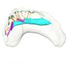
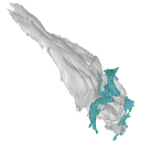
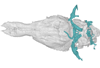
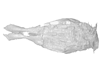
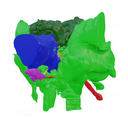
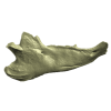
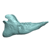
The present 3D Dataset contains the 3D models analyzed in Mennecart, B., Duranthon, F., & Costeur, L. 2024. Systematic contribution of the auditory region to the knowledge of the oldest European Bovidae (Mammalia, Ruminantia). Journal of Anatomy XXX. https://doi.org/10.1111/joa.14132
Pusillutragus montrealensis MHNT.PAL.2015.0.2261.4 View specimen
|
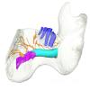
M3#1522Right petrosal, bony labyrinth, stapes Type: "3D_surfaces"doi: 10.18563/m3.sf.1522 state:published |
Download 3D surface file |
Pusillutragus montrealensis MHNT.PAL.2015.0.2261.9 View specimen
|
M3#1523Left petrosal and left bony labyrinth Type: "3D_surfaces"doi: 10.18563/m3.sf.1523 state:published |
Download 3D surface file |
Eotragus artenensis SMNS-P-41625 View specimen

|
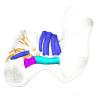
M3#1524Petrosal (right), bony labyrinth (left) Type: "3D_surfaces"doi: 10.18563/m3.sf.1524 state:published |
Download 3D surface file |
Eotragus clavatus NMB San.15056 View specimen
|
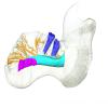
M3#1528Right petrosal and right bony labyrinth Type: "3D_surfaces"doi: 10.18563/m3.sf.1528 state:published |
Download 3D surface file |
Eotragus clavatus NMB San.15055 View specimen
|
M3#1526Left Petrosal Type: "3D_surfaces"doi: 10.18563/m3.sf.1526 state:published |
Download 3D surface file |
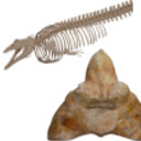
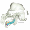
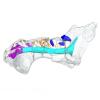
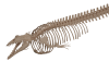
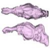
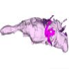
The present 3D Dataset contains the 3D models analyzed in Bianucci et al. 2023, A heavyweight early whale pushes the boundaries of vertebrate morphology, Nature. These include bones of the holotype of new species Perucetus colossus (MUSM 3248), as well as the articulated skeleton of Cynthiacetus peruvianus (holotype, MNHN.F.PRU10). The latter was used to estimate the total skeleton volume of P. colossus.
Perucetus colossus MUSM 3248 View specimen
|
M3#1131Thirteen vertebrae, rib, and innominate of Perucetus colossus (holotype, MUSM NNNN). Type: "3D_surfaces"doi: 10.18563/m3.sf.1131 state:published |
Download 3D surface file |
Cynthiacetus peruvianus MNHN.F.PRU10 View specimen
|
M3#1130Articulated skeleton of the holotype of Cynthiacetus peruvianus MNHN.F.PRU10 Type: "3D_surfaces"doi: 10.18563/m3.sf.1130 state:published |
Download 3D surface file |
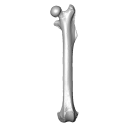
This 3D Dataset includes the 3D models analysed in Wölfer J & Hautier L. 2024 Inferring the locomotor ecology of two of the oldest fossil squirrels: influence of operationalisation, trait, body size, and machine learning method. Proceedings of the Royal Society B. https://doi.org/10.1098/rspb.2024-0743
Palaeosciurus goti MGB125 View specimen

|
M3#1577Left femur of Palaeosciurus goti Type: "3D_surfaces"doi: 10.18563/m3.sf.1577 state:published |
Download 3D surface file |
Palaeosciurus feignouxi GER291 View specimen
|
M3#1578Right femur of Palaeosciurus feignouxi Type: "3D_surfaces"doi: 10.18563/m3.sf.1578 state:published |
Download 3D surface file |
Palaeosciurus feignouxi GER293 View specimen
|
M3#1579Right femur of Palaeosciurus feignouxi Type: "3D_surfaces"doi: 10.18563/m3.sf.1579 state:published |
Download 3D surface file |
Palaeosciurus feignouxi GER294 View specimen
|
M3#1580Right femur of Palaeosciurus feignouxi Type: "3D_surfaces"doi: 10.18563/m3.sf.1580 state:published |
Download 3D surface file |
Palaeosciurus feignouxi GER296 View specimen
|
M3#1581Left femur of Palaeosciurus feignouxi Type: "3D_surfaces"doi: 10.18563/m3.sf.1581 state:published |
Download 3D surface file |
Palaeosciurus feignouxi GER298 View specimen
|
M3#1582Left femur of Palaeosciurus feignouxi Type: "3D_surfaces"doi: 10.18563/m3.sf.1582 state:published |
Download 3D surface file |
Palaeosciurus feignouxi GER299 View specimen
|
M3#1583Left femur of Palaeosciurus feignouxi Type: "3D_surfaces"doi: 10.18563/m3.sf.1583 state:published |
Download 3D surface file |
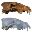
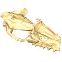
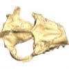
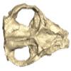
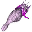
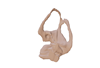
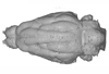
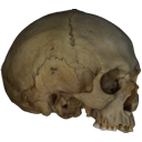
Speothos pacivorus is an extinct South American canid (Canidae: Cerdocyonina) from the Pleistocene of Lagoa Santa Karst, Central Brazil. This taxon is one of the hypercarnivore canids that vanished from the continent at the end of Pleistocene. Although all remains of Speothos pacivorus were collected in the 19th century by the Danish naturalist Peter W. Lund, few studies have committed to an in-depth analysis of the taxon and the known specimens. Here, we analyzed all biological remains of S. pacivorus hosted in the Peter Lund/Quaternary Collection at the Natural History Museum of Denmark, Copenhagen, by listing and illustrating all its specimens known to date. We also conducted a reconstruction of the holotype, an almost complete cranium, based on a µCT scan, producing an undeformed and crack-free three-dimensional model. With this data available we aim to foster new research on this elusive species.
Speothos pacivorus NHMD:211341 View specimen
|
M3#1475Holotype of Speothos pacivorus Type: "3D_surfaces"doi: 10.18563/m3.sf.1475 state:published |
Download 3D surface file |
The present 3D Dataset contains the 3D model of the skin of Allosaurus described in Hendrickx, C. et al. in press. Morphology and distribution of scales, dermal ossifications, and other non-feather integumentary structures in non-avialan theropod dinosaurs. Biological Reviews.
Allosaurus jimmadseni UMNH VP C481 View specimen
|
M3#902The material consists of a 3D reconstruction of the counterpart of a 30 cm2 patch of skin impression associated with the anterior dorsal ribs/pectoral region of the specimen of Allosaurus jimmadseni UMNH VP C481. The skin shows a semi-uniform basement of 1-2 mm diameter pebbles with a smaller number of slightly larger (up to 3 mm) ovoid scales. The irregular shape, distribution, and overall small size of these larger scales suggest that they are not classifiable as feature scales but rather as variations in the basement scales. Type: "3D_surfaces"doi: 10.18563/m3.sf.902 state:published |
Download 3D surface file |
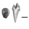
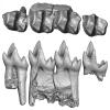
The present 3D Dataset contains the 3D model of the skull of the raoellid Indohyus indirae described in Patel et al. 2024.
Indohyus indirae RR 207 View specimen
|
M3#1259dorsoventrally crushed skull Type: "3D_surfaces"doi: 10.18563/m3.sf.1259 state:published |
Download 3D surface file |
Indohyus indirae RR 601 View specimen
|
M3#1268dorsoventrally crushed skull Type: "3D_surfaces"doi: 10.18563/m3.sf.1268 state:published |
Download 3D surface file |
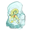
The present 3D Dataset contains the 3D models of the endocranial cast of two specimens of Indohyus indirae described in the article entitled “The endocranial cast of Indohyus (Artiodactyla, Raoellidae): the origin of the cetacean brain” (Orliac and Thewissen, 2021). They represent the cast of the main cavity of the braincase as well as associated intraosseous sinuses.
Indohyus indirae RR 207 View specimen
|
M3#710cast of the main endocranial cavity and associated intraosseous sinuses Type: "3D_surfaces"doi: 10.18563/m3.sf.710 state:published |
Download 3D surface file |
Indohyus indirae RR 601 View specimen
|
M3#711casts of the main endocranial cavity Type: "3D_surfaces"doi: 10.18563/m3.sf.711 state:published |
Download 3D surface file |
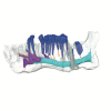
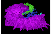
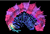
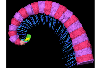
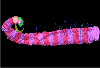
Current knowledge on the skeletogenesis of Chondrichthyes is scarce compared with their extant sister group, the bony fishes. Most of the previously described developmental tables in Chondrichthyes have focused on embryonic external morphology only. Due to its small body size and relative simplicity to raise eggs in laboratory conditions, the small-spotted catshark Scyliorhinus canicula has emerged as a reference species to describe developmental mechanisms in the Chondrichthyes lineage. Here we investigate the dynamic of mineralization in a set of six embryonic specimens using X-ray microtomography and describe the developing units of both the dermal skeleton (teeth and dermal scales) and endoskeleton (vertebral axis). This preliminary data on skeletogenesis in the catshark sets the first bases to a more complete investigation of the skeletal developmental in Chondrichthyes. It should provide comparison points with data known in osteichthyans and could thus be used in the broader context of gnathostome skeletal evolution.
Scyliorhinus canicula SC6_2_2015_03_20 View specimen

|
M3#50Mineralized skeleton of a 6,2 cm long embryo of Scyliorhinus canicula Type: "3D_surfaces"doi: 10.18563/m3.sf.50 state:published |
Download 3D surface file |
Scyliorhinus canicula SC6_7_2015_03_20 View specimen

|
M3#51Mineralized skeleton of a 6,7 cm long embryo of Scyliorhinus canicula Type: "3D_surfaces"doi: 10.18563/m3.sf.51 state:published |
Download 3D surface file |
Scyliorhinus canicula SC7_1_2015_04_03 View specimen

|
M3#52Mineralized skeleton of a 7,1 cm long embryo of Scyliorhinus canicula Type: "3D_surfaces"doi: 10.18563/m3.sf.52 state:published |
Download 3D surface file |
Scyliorhinus canicula SC7_5_2015_03_13 View specimen

|
M3#53Mineralized skeleton of a 7,5 cm long embryo of Scyliorhinus canicula Type: "3D_surfaces"doi: 10.18563/m3.sf.53 state:published |
Download 3D surface file |
Scyliorhinus canicula SC8_2015_03_20 View specimen

|
M3#54Mineralized skeleton of a 8 cm long embryo of Scyliorhinus canicula Type: "3D_surfaces"doi: 10.18563/m3.sf.54 state:published |
Download 3D surface file |
Scyliorhinus canicula SC10_2015_02_27 View specimen

|
M3#55Mineralized skeleton of a 10 cm long embryo of Scyliorhinus canicula Type: "3D_surfaces"doi: 10.18563/m3.sf.55 state:published |
Download 3D surface file |
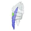
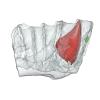
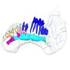
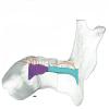
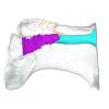
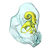
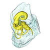
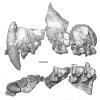
The present 3D dataset contains 3D models of the endocranial cast of the raoellid Khirtharia inflata retrieved from the middle Eocene of the Upper Subathu Formation in the Kalakot area (India). Raoellidae are closely related to stem cetaceans and bring crucial information to understand the earliest phase of land to water transition in Cetacea.
Khirtharia inflata GU/RJ/197 View specimen
|
M3#1608labeled cast of the endocranial cavity Type: "3D_surfaces"doi: 10.18563/m3.sf.1608 state:published |
Download 3D surface file |
|
M3#1609endocast and associated sinuses Type: "3D_surfaces"doi: 10.18563/m3.sf.1609 state:published |
Download 3D surface file |
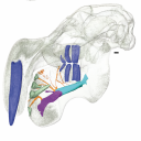
The present 3D Dataset contains the 3D models analyzed in Pochat-Cottilloux Y., Martin J.E., Jouve S., Perrichon G., Adrien J., Salaviale C., de Muizon C., Cespedes R. & Amiot R. (2021). The neuroanatomy of Zulmasuchus querejazus (Crocodylomorpha, Sebecidae) and its implications for the paleoecology of sebecosuchians. The Anatomical Record, https://doi.org/10.1002/ar.24826
Zulmasuchus querejazus MHNC 6672 View specimen
|
M3#798Left endosseous labyrinth of Z. querejazus (MHNC 6672). Type: "3D_surfaces"doi: 10.18563/m3.sf.798 state:published |
Download 3D surface file |
|
M3#799Reconstruction of the endocranial cavities of Z. querejazus (MHNC 6672). Type: "3D_surfaces"doi: 10.18563/m3.sf.799 state:published |
Download 3D surface file |
|
M3#800Three-dimensional reconstruction of the pneumatic cavities within the braincase of Z. querejazus (MHNC 6672) Type: "3D_surfaces"doi: 10.18563/m3.sf.800 state:published |
Download 3D surface file |
Our knowledge of the external brain morphology of the late Eocene artiodactyl ungulate Mixtotherium, relies on a plaster model realized on a specimen from the Victor Brun Museum in Montauban (France) and described by Dechaseaux (1973). Here, based on micro CT-scan data, we virtually reconstruct the 3D cast of the empty cavity of the partial cranium MA PHQ 716 from the Victor Brun Museum and compare it to the plaster model illustrated and described by Dechaseaux (1973). Indeed, the specimen from which the original plaster endocast originates was not identified by Dechaseaux by a specimen number. We confirm here that the studied specimen was indeed the one described and illustrated by Dechaseaux (1973). We also reconstruct a second, more detailed, model providing additional morphological and quantitative observations made available by micro CT scan investigation such as precisions on the neopallium folding and endocranial volumes.
Mixtotherium cuspidatum MA PHQ 716 View specimen
|
M3#857endocast of the brain cavity Type: "3D_surfaces"doi: 10.18563/m3.sf.857 state:published |
Download 3D surface file |
The present 3D Dataset contains the 3D models of the skull of the holotype of Miocaperea pulchra.
Miocaperea pulchra SMNS-P-46978 View specimen
|
M3#1656Blender file containing two models (the skull being preserved in two parts) Type: "3D_surfaces"doi: 10.18563/m3.sf.1656 state:published |
Download 3D surface file |
This contribution contains the three-dimensional models of the most complete and/or informative fossil materials attributed to Peradectes crocheti Gernelle, 2024, the earliest peradectid metatherian species of Europe, from its type locality (Palette, Provence, ~55 Ma). These specimens were analyzed and discussed in: Gernelle et al. (2024), Taxonomy and evolutionary history of peradectids (Metatheria): new data from the early Eocene of France. https://doi.org/10.1007/s10914-024-09724-5
Peradectes crocheti MHN.AIX.PV.2018.26.14 View specimen
|
M3#14993D surface model of MHN.AIX.PV.2018.26.14, fragmentary left maxilla with C-P1, anterior root of P2, and M1-M3 Type: "3D_surfaces"doi: 10.18563/m3.sf.1499 state:published |
Download 3D surface file |
Peradectes crocheti MHN.AIX.PV.2017.6.6 View specimen
|
M3#15003D surface model of MHN.AIX.PV.2017.6.6, left P2 Type: "3D_surfaces"doi: 10.18563/m3.sf.1500 state:published |
Download 3D surface file |
Peradectes crocheti MHN.AIX.PV.2017.6.7 View specimen
|
M3#15013D surface model of MHN.AIX.PV.2017.6.7, left M3 Type: "3D_surfaces"doi: 10.18563/m3.sf.1501 state:published |
Download 3D surface file |
Peradectes crocheti MHN.AIX.PV.2017.6.8 View specimen
|
M3#15023D surface model of MHN.AIX.PV.2017.6.8, right hemi-mandible fragment with canine alveolus, posterior root of p1, partial p2, p3, partial m1, and m2-m3 Type: "3D_surfaces"doi: 10.18563/m3.sf.1502 state:published |
Download 3D surface file |
Peradectes crocheti MHN.AIX.PV.2017.6.9 View specimen
|
M3#15033D surface model of MHN.AIX.PV.2017.6.9, leftm1-m4 row with fragments of dentary Type: "3D_surfaces"doi: 10.18563/m3.sf.1503 state:published |
Download 3D surface file |
Peradectes crocheti MHN.AIX.PV.2017.6.14 View specimen
|
M3#15043D surface model of MHN.AIX.PV.2017.6.14, right astragalus Type: "3D_surfaces"doi: 10.18563/m3.sf.1504 state:published |
Download 3D surface file |
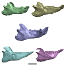
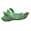
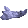
The present 3D Dataset contains the 3D models analyzed in 3D Finite Element Analysis and Geometric Morphometrics of Sloths (Xenarthra, Folivora) Mandibles Show Insights on the Dietary Specializations of Fossil Taxa. Journal of South American Earth Sciences. https://doi.org/10.1016/j.jsames.2023.104445
Mylodon darwinii CAV 379 View specimen
|
M3#1159Right hemimandible Type: "3D_surfaces"doi: 10.18563/m3.sf.1159 state:published |
Download 3D surface file |
Scelidotherium leptocephalum MNHN-M 137,722 View specimen
|
M3#1160Mandible Type: "3D_surfaces"doi: 10.18563/m3.sf.1160 state:published |
Download 3D surface file |
Glossotherium robustum MNHN-M 914 View specimen
|
M3#1161Mandible Type: "3D_surfaces"doi: 10.18563/m3.sf.1161 state:published |
Download 3D surface file |
Lestodon armatus MPAC 899 View specimen
|
M3#1162Mandible Type: "3D_surfaces"doi: 10.18563/m3.sf.1162 state:published |
Download 3D surface file |
Valgipes bucklandi NHMD.Z.M.K. 1/1845:3540 View specimen
|
M3#1163Mandible Type: "3D_surfaces"doi: 10.18563/m3.sf.1163 state:published |
Download 3D surface file |
The present 3D Dataset contains the 3D models illustrated and described in the chapter “Paleoneurology of Artiodactyla, an overview of the evolution of the artiodactyl brain” (Orliac et al. 2022) published in "Paleoneurology of amniotes: new directions in the study of fossil endocasts", edited by Dozo, Paulina-Carabajal, Macrini and Walsh.
Homacodon vagans AMNH 12695 View specimen
|
M3#1063Endocranial cast Type: "3D_surfaces"doi: 10.18563/m3.sf.1063 state:published |
Download 3D surface file |
Helohyus sp. AMNH 13079 View specimen
|
M3#1064Endocranial cast Type: "3D_surfaces"doi: 10.18563/m3.sf.1064 state:published |
Download 3D surface file |
Leptauchenia sp. AMNH 45508 View specimen
|
M3#1065endocranial cast Type: "3D_surfaces"doi: 10.18563/m3.sf.1065 state:published |
Download 3D surface file |
Agriochoerus sp. AMNH 95330 View specimen
|
M3#1067endocranial cast Type: "3D_surfaces"doi: 10.18563/m3.sf.1067 state:published |
Download 3D surface file |
Mouillacitherium elegans UM ACQ 6625 View specimen
|
M3#1068endocranial cast Type: "3D_surfaces"doi: 10.18563/m3.sf.1068 state:published |
Download 3D surface file |
Caenomeryx filholi UM PDS 2570 View specimen
|
M3#1069endocranial cast Type: "3D_surfaces"doi: 10.18563/m3.sf.1069 state:published |
Download 3D surface file |
Dichobune leporina MNHN.F.QU16586 View specimen
|
M3#1070endocranial cast Type: "3D_surfaces"doi: 10.18563/m3.sf.1070 state:published |
Download 3D surface file |
Anoplotherium sp. not numbered View specimen
|
M3#1071endocranial cast Type: "3D_surfaces"doi: 10.18563/m3.sf.1071 state:published |
Download 3D surface file |
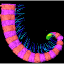
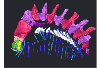
The present 3D Dataset contains the 3D models analyzed in the publication Fossils from the Montceau-les-Mines Lagerstätte (305 Ma) shed light on the anatomy, ecology and phylogeny of Carboniferous millipedes. Authors: Lheritier Mickael, Perroux Maëva, Vannier Jean, Escarguel Gilles, Wesener Thomas, Moritz Leif, Chabard Dominique, Adrien Jerome and Perrier Vincent. Journal of Systematics Palaeontology. https://doi.org/10.1080/14772019.2023.2169891
Amynilyspes fatimae MNHN.F.SOT.2134 View specimen
|
M3#1073Nearly complete specimen. Type: "3D_surfaces"doi: 10.18563/m3.sf.1073 state:published |
Download 3D surface file |
Amynilyspes fatimae MNHN.F.SOT.14983 View specimen
|
M3#1074Nearly complete specimen. Type: "3D_surfaces"doi: 10.18563/m3.sf.1074 state:published |
Download 3D surface file |
Amynilyspes fatimae MNHN.F.SOT.2129 View specimen
|
M3#1075Nearly complete specimen. Type: "3D_surfaces"doi: 10.18563/m3.sf.1075 state:published |
Download 3D surface file |
Blanzilius parriati MNHN.F.SOT.2114A View specimen
|
M3#1076Front part. Type: "3D_surfaces"doi: 10.18563/m3.sf.1076 state:published |
Download 3D surface file |
Blanzilius parriati MNHN.F.SOT.5148 View specimen
|
M3#1077Front part. Type: "3D_surfaces"doi: 10.18563/m3.sf.1077 state:published |
Download 3D surface file |
Blanzilius parriati MNHN.F.SOT.2113 View specimen
|
M3#1078Fragment with legs, sternites and possible tracheal openings. Type: "3D_surfaces"doi: 10.18563/m3.sf.1078 state:published |
Download 3D surface file |
Blanzilius parriati MNHN.F.SOT.81522 View specimen
|
M3#1079Nealry complete specimen. Type: "3D_surfaces"doi: 10.18563/m3.sf.1079 state:published |
Download 3D surface file |
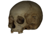
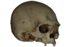
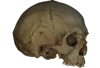
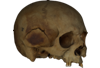
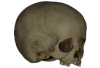
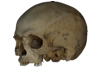
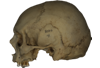
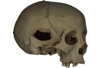
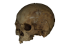
This contribution contains the 3D models described and figured in the following publications:
- Marini E., Lussu P., 2020. A virtual physical anthropology lab. Teaching in the time of coronavirus, in prep.;
- Lussu P., Bratzu D., Marini E., 2020. Cloud-based ultra close-range digital photogrammetry: validation of an approach for the effective virtual reconstruction of skeletal remains, in prep.
Homo sapiens MSAE 59 View specimen

|
M3#509MSAE 59 Type: "3D_surfaces"doi: 10.18563/m3.sf.509 state:published |
Download 3D surface file |
Homo sapiens MSAE 62 View specimen
|
M3#510MSAE 62 Type: "3D_surfaces"doi: 10.18563/m3.sf.510 state:published |
Download 3D surface file |
Homo sapiens MSAE 63 View specimen
|
M3#512MSAE 63 Type: "3D_surfaces"doi: 10.18563/m3.sf.512 state:published |
Download 3D surface file |
Homo sapiens MSAE 78 View specimen
|
M3#514MSAE 78 Type: "3D_surfaces"doi: 10.18563/m3.sf.514 state:published |
Download 3D surface file |
Homo sapiens MSAE 95 View specimen
|
M3#515MSAE 95 Type: "3D_surfaces"doi: 10.18563/m3.sf.515 state:published |
Download 3D surface file |
Homo sapiens MSAE 1852 View specimen
|
M3#516MSAE 1852 Type: "3D_surfaces"doi: 10.18563/m3.sf.516 state:published |
Download 3D surface file |
Homo sapiens MSAE 6426 View specimen
|
M3#517MSAE 6426 Type: "3D_surfaces"doi: 10.18563/m3.sf.517 state:published |
Download 3D surface file |
Homo sapiens MSAE 6428 View specimen
|
M3#518MSAE 6428 Type: "3D_surfaces"doi: 10.18563/m3.sf.518 state:published |
Download 3D surface file |
Homo sapiens MSAE 6992 View specimen
|
M3#519MSAE 6992 Type: "3D_surfaces"doi: 10.18563/m3.sf.519 state:published |
Download 3D surface file |
Homo sapiens MSAE 7688 View specimen
|
M3#520MSAE 7688 Type: "3D_surfaces"doi: 10.18563/m3.sf.520 state:published |
Download 3D surface file |


