Explodable 3D Dog Skull for Veterinary Education
3D models of Ocnotherium skull
3D models of Kalakocetus, the earliest Cetacea
3D GM dataset of bird skeletal variation
Skeletal embryonic development in the catshark
Bony connexions of the petrosal bone of extant hippos
bony labyrinth (14) , inner ear (11) , geometric morphometrics (10) , CT-scan (10) , Eocene (10) , Micro-CT (9) , Miocene (8)
Lionel Hautier (24) , Maëva Judith Orliac (23) , Laurent Marivaux (18) , Renaud Lebrun (14) , Rodolphe Tabuce (14) , Pierre-Olivier Antoine (13) , Bastien Mennecart (13)
|
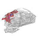
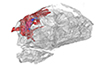
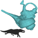
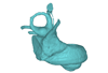
3D models related to the publication: New information on the braincase and endocranial morphology of the Late Triassic neotheropod Zupaysaurus rougieri using Computed Tomography dataAriana Paulina-Carabajal
Published online: 26/08/2019 |
|
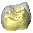
M3#424The Zip contains 3 files, which correspond to: PULR_076-M1: Zupaysaurus rougieri skull, braincase and cranial endocast PULR_076-M2: Zupaysaurus rougieri braincase PULR_076-M1: Zupaysaurus rougieri brain and inner ear Type: "3D_surfaces"doi: 10.18563/m3.sf.424 state:published |
Download 3D surface file |
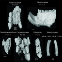
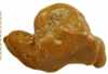
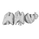
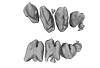
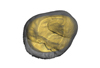
This contribution contains 3D models of extinct rodents Dinomyidae from Miocene and Quaternary of Brazil. The Miocene specimens that were digitalized include the holotypes of Potamarchus adamiae, Pseudopotamarchus villanuevai, and Ferigolomys pacarana collected in the Solimões Formation (Upper Miocene), northern Brazil. The Quaternary specimens are the holotype and paratype of Niedemys piauiensis, found in Upper Pleistocene deposits from northeast Brazil.
Potamarchus adamiae UFAC-CS 011 View specimen
|
M3#410UFAC-CS 011 – holotype, palatal region of the skull with cheek teeth Type: "3D_surfaces"doi: 10.18563/m3.sf.410 state:published |
Download 3D surface file |
Potamarchus adamiae UFAC-CS 043 View specimen
|
M3#411UFAC-CS 043, left dentary with cheek teeth Type: "3D_surfaces"doi: 10.18563/m3.sf.411 state:published |
Download 3D surface file |
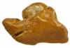
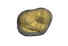
Pseudopotamarchus villanuevai UFAC 4762 View specimen
|
M3#412UFAC 4762 – holotype, incomplete right maxilla with cheek teeth Type: "3D_surfaces"doi: 10.18563/m3.sf.412 state:published |
Download 3D surface file |
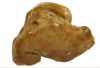
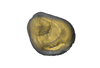
Ferigolomys pacarana UFAC 6460 View specimen

|
M3#413UFAC 6460 – holotype, palatal region of the skull with cheek teeth Type: "3D_surfaces"doi: 10.18563/m3.sf.413 state:published |
Download 3D surface file |
Drytomomys sp. UFAC 2742 View specimen
|
M3#414UFAC 2742, right dentary with cheek teeth Type: "3D_surfaces"doi: 10.18563/m3.sf.414 state:published |
Download 3D surface file |
Niedemys piauiensis FUMDHAM 113-146365-2 View specimen
|
M3#418FUMDHAM 113-146365-2 - holotype, upper right tooth Type: "3D_surfaces"doi: 10.18563/m3.sf.418 state:published |
Download 3D surface file |
Niedemys piauiensis FUMDHAM 113-145304-2 View specimen
|
M3#419FUMDHAM 113-145304-2 - paratype, left lower molar Type: "3D_surfaces"doi: 10.18563/m3.sf.419 state:published |
Download 3D surface file |
This contribution contains the 3D model described and figured in the following publication: New turtles from the Late Cretaceous of Monte Alto-SP, Brazil, including cranial osteology, neuroanatomy and phylogenetic position of a new taxon. PalZ. https://doi.org/10.1007/s12542-017-0397-x
Yuraramirim montealtensis 04-0008/89 View specimen
|
M3#2783D surfaces related to specimen MPMA 04-0008/89. Type: "3D_surfaces"doi: 10.18563/m3.sf.278 state:published |
Download 3D surface file |
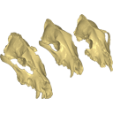
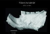
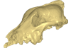
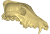
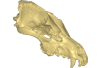
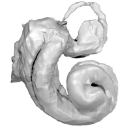
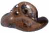
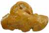
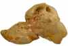
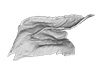
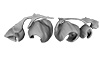
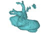
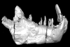
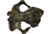
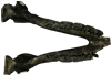
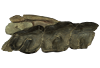
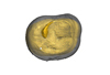
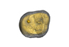
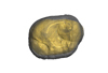
Archaeozoological studies are increasingly using new methods and approaches to explore questions about domestication. Here, we provide 3D models of three archaeological Canis lupus skulls from Belgium originating from the sites of Goyet (31,680±250BP; 31,890+240/-220BP), Trou des Nutons (21,810±90BP) and Trou Balleux (postglacial). Since their identification as either wolves or early dogs is still debated, we present these models as additional tools for further investigating their evolutionary history and the history of dog domestication.
Canis lupus Goyet 2860 View specimen
|
M3#213D surface model of the cranium of the Late Pleistocene Canis lupus "Goyet 2860" from the Royal Belgian Institute of Natural Sciences. Type: "3D_surfaces"doi: 10.18563/m3.sf21 state:published |
Download 3D surface file |
Canis lupus Trou Balleux no-nr View specimen
|
M3#223D surface model of the cranium of the Late Pleistocene Canis lupus "Trou Balleux no-nr" from the University of Liège, Belgium Type: "3D_surfaces"doi: 10.18563/m3.sf22 state:published |
Download 3D surface file |
Canis lupus Trou des Nutons 2559-1 View specimen
|
M3#233D surface model of the cranium of the Late Pleistocene Canis lupus "Trou des Nutons 2559-1" from the Royal Belgian Institute of Natural Sciences. Type: "3D_surfaces"doi: 10.18563/m3.sf23 state:published |
Download 3D surface file |
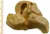
This contribution contains the 3D reconstruction of Canariomys bravoi, described and figured in the following publication: Michaux J., Hautier L., Hutterer R., Lebrun R., Guy F., García-Talavera F., 2012 : Body shape and life style of the extinct rodent Canariomys bravoi (Mammalia, Murinae) from Tenerife, Canary Islands (Spain). Comptes Rendus Palevol 11 (7), 485-494. DOI: 10.1016/j.crpv.2012.06.004
Canariomys bravoi TFMCV872-873 View specimen

|
M3#6This file contains the 3D reconstruction of Canariomys bravoi, described and figured in the following publication: Michaux J., Hautier L., Hutterer R., Lebrun R., Guy F., García-Talavera F., 2012 : Body shape and life style of the extinct rodent Canariomys bravoi (Mammalia, Murinae) from Tenerife, Canary Islands (Spain). Comptes Rendus Palevol 11 (7), 485-494. Type: "3D_surfaces"doi: 10.18563/m3.sf6 state:published |
Download 3D surface file |
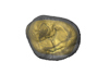
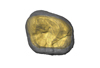
This contribution contains the 3D models described and figured in the following publication: Aguirre-Fernández G, Jost J, and Hilfiker S. 2022. First records of extinct kentriodontid and squalodelphinid dolphins from the Upper Marine Molasse (Burdigalian age) of Switzerland and a reappraisal of the Swiss cetacean fauna.
Kentriodon sp. NMBE 5023944 View specimen
|
M3#8583D models of left periotic and bony labyrinth of NMBE 5023944 (Kentriodon sp.) Type: "3D_surfaces"doi: 10.18563/m3.sf.858 state:published |
Download 3D surface file |
Kentriodon sp. NMBE 5023945 View specimen
|
M3#8593D models of right periotic and bony labyrinth of NMBE 5023945 (Kentriodontidae indet.) Type: "3D_surfaces"doi: 10.18563/m3.sf.859 state:published |
Download 3D surface file |
Kentriodon sp. NMBE 5023946 View specimen
|
M3#8603D models of left periotic and bony labyrinth of NMBE 5023946 (Kentriodon sp.) Type: "3D_surfaces"doi: 10.18563/m3.sf.860 state:published |
Download 3D surface file |
Kentriodon sp. NMBE 5036436 View specimen
|
M3#8613D models of right periotic and bony labyrinth of NMBE 5036436 (Kentriodontidae indet.) Type: "3D_surfaces"doi: 10.18563/m3.sf.861 state:published |
Download 3D surface file |
indet. indet. NMBE 5023942 View specimen
|
M3#8623D models of right periotic and bony labyrinth of NMBE 5023942 (Squalodelphinidae indet.) Type: "3D_surfaces"doi: 10.18563/m3.sf.862 state:published |
Download 3D surface file |
indet. indet. NMBE 5023943 View specimen
|
M3#8633D models of left periotic and bony labyrinth of NMBE 5023943 (Squalodelphinidae indet.) Type: "3D_surfaces"doi: 10.18563/m3.sf.863 state:published |
Download 3D surface file |
indet. indet. NMBE 5036437 View specimen
|
M3#8643D models of left periotic and bony labyrinth of NMBE 5036437 (Physeteridae indet.) Type: "3D_surfaces"doi: 10.18563/m3.sf.864 state:published |
Download 3D surface file |
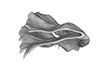
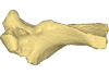
The present 3D Dataset contains the 3D model analyzed in Presence of the ground sloth Valgipes bucklandi (Xenarthra, Folivora, Scelidotheriinae) in southern Uruguay during the Late Pleistocene: Ecological and biogeographical implications. Quaternary International. https://doi.org/10.1016/j.quaint.2021.06.011
Valgipes bucklandi CAV 1573 View specimen
|
M3#797Left tibia-fibula Type: "3D_surfaces"doi: 10.18563/m3.sf.797 state:published |
Download 3D surface file |
The present 3D Dataset contains the 3D models analyzed in ”Morphological features of tooth development and replacement in the rabbit Oryctolagus cuniculus”, Archives of Oral Biology, https://doi.org/10.1016/j.archoralbio.2019.104576
Oryctogalus cuniculus E14 View specimen
|
M3#390Right cheek teeth, Left and right incisors at 14 dpf Type: "3D_surfaces"doi: 10.18563/m3.sf.390 state:published |
Download 3D surface file |
Oryctogalus cuniculus E16 View specimen
|
M3#391Left cheek teeth, Left and right incisors at 16 dpf Type: "3D_surfaces"doi: 10.18563/m3.sf.391 state:published |
Download 3D surface file |
Oryctogalus cuniculus E18 View specimen
|
M3#392Left cheek teeth and incisors at 18 dpf Type: "3D_surfaces"doi: 10.18563/m3.sf.392 state:published |
Download 3D surface file |
Oryctogalus cuniculus E20 View specimen
|
M3#393Left cheek teeth and incisors at 20 dpf Type: "3D_surfaces"doi: 10.18563/m3.sf.393 state:published |
Download 3D surface file |
Oryctogalus cuniculus E22 View specimen
|
M3#394Left lower cheek teeth and incisors, right upper cheek teeth and incisors at 22 dpf Type: "3D_surfaces"doi: 10.18563/m3.sf.394 state:published |
Download 3D surface file |
Oryctogalus cuniculus E24 View specimen
|
M3#395Left cheek teeth and incisors at 24 dpf Type: "3D_surfaces"doi: 10.18563/m3.sf.395 state:published |
Download 3D surface file |
Oryctogalus cuniculus E28 View specimen
|
M3#396Right cheek teeth and incisors at 28 dpf Type: "3D_surfaces"doi: 10.18563/m3.sf.396 state:published |
Download 3D surface file |
Oryctogalus cuniculus E26 View specimen
|
M3#397Right cheek teeth and incisors at 26 dpf Type: "3D_surfaces"doi: 10.18563/m3.sf.397 state:published |
Download 3D surface file |
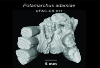
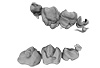
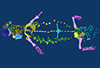
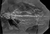
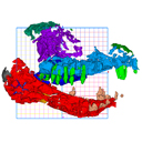
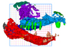
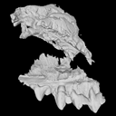
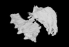
The present contribution contains the 3D model and dataset analyzed in the following publication: Scheyer, T. M., J. M. Neenan, T. Bodogan, H. Furrer, C. Obrist, and M. Plamondon. 2017. A new, exceptionally preserved juvenile specimen of Eusaurosphargis dalsassoi (Diapsida) and implications for Mesozoic marine diapsid phylogeny. Scientific Reports, https://doi.org/10.1038/s41598-017-04514-x .
Eusaurosphargis dalsassoi PIMUZ A/III 4380 View specimen
|
M3#17994 extracted surfaces of skeletal elements of PIMUZ A/III 4380 Type: "3D_surfaces"doi: 10.18563/m3.sf.179 state:published |
Download 3D surface file |
|
M3#180Accompanying CT scan dataset Type: "3D_CT"doi: 10.18563/m3.sf.180 state:published |
Download CT data |
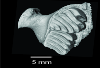
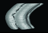
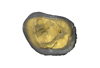
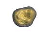
This contribution contains the 3D models of the bony labyrinths of two protocetid archaeocetes from the locality of Kpogamé, Togo, described and figured in the publication of Mourlam and Orliac (2017). https://doi.org/10.1016/j.cub.2017.04.061
?Carolinacetus indet. UM KPG-M 164 View specimen
|
M3#149bony labyrinth of ? Carolinacetus sp. from Kpogamé, Togo Type: "3D_surfaces"doi: 10.18563/m3.sf.149 state:published |
Download 3D surface file |
indet. indet. UM KPG-M 73 View specimen
|
M3#150bony labyrinth of Protocetidae indet. from Kpogamé, Togo Type: "3D_surfaces"doi: 10.18563/m3.sf.150 state:published |
Download 3D surface file |
The present 3D Dataset contains the 3D model analyzed in Gaetano, L. C., Abdala, F., Seoane, F. D., Tartaglione, A., Schulz, M., Otero, A., Leardi, J. M., Apaldetti, C., Krapovickas, V., and Steinbach, E. 2021. A new cynodont from the Upper Triassic Los Colorados Formation (Argentina, South America) reveals a novel paleobiogeographic context for mammalian ancestors. Scientific Reports.
Tessellatia bonapartei PULR-V121 View specimen
|
M3#9603D surface model of PULR-V121 Type: "3D_surfaces"doi: 10.18563/m3.sf.960 state:published |
Download 3D surface file |
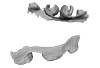
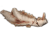
This contribution contains the 3D model(s) described and figured in the following publication: The present 3D Dataset contains the 3D models and CT-Scan slices of the lower jaws and teeth analyzed in “A new prozostrodontian cynodont (Eucynodontia, Probainognathia) from the Upper Triassic of southern Brazil”. https://doi.org/10.1080/02724634.2020.1782415
Agudotherium gassenae CAPPA/UFSM 0262 View specimen
|
M3#546Left lower jaw and cheek teeth Type: "3D_surfaces"doi: 10.18563/m3.sf.546 state:published |
Download 3D surface file |
|
M3#5471578 slices Type: "3D_CT"doi: 10.18563/m3.sf.547 state:published |
Download CT data |
Agudotherium gassenae CAPPA/UFSM 0208 View specimen
|
M3#548right lower jaw Type: "3D_surfaces"doi: 10.18563/m3.sf.548 state:published |
Download 3D surface file |
|
M3#549CT data of CAPPA_UFSM_0208 Type: "3D_CT"doi: 10.18563/m3.sf.549 state:published |
Download CT data |
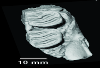
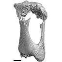
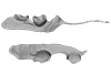
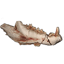
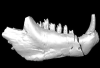
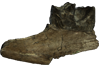
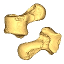
The present 3D Dataset contains the 3D model analyzed in the following publication: Solé et al. (2018), Niche partitioning of the European carnivorous mammals during the paleogene. Palaios. https://doi.org/10.2110/palo.2018.022
Hyaenodon leptorhynchus FSL848325 View specimen
|
M3#336The specimen FSL848325 is separated in two fragments: the anterior part bears the incisors, the deciduous and permanent canines, while the posterior part bears the right P3, P4, M1 and M2. The P2 is isolated. When combined, the cranium length is approximatively 10.5 cm long. The anterior part is 6.9 cm long and 2.15 cm wide (taken at the level of the P1). The posterior part is 4.8 cm long. The anterior part of the cranium is very narrow. Type: "3D_surfaces"doi: 10.18563/m3.sf.336 state:published |
Download 3D surface file |
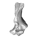
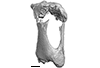
This contribution contains the 3D model described and figured in the following publication: Crochet, J.-Y., Hautier, L., Lehmann, T., 2015. A pangolin (Manidae, Pholidota, Mammalia) from the French Quercy phosphorites (Pech du Fraysse, Saint-Projet, Tarn-et-Garonne, late Oligocene, MP 28). Palaeovertebrata 39(2)-e4. doi: 10.18563/pv.39.2.e4
Necromanis franconica UM PFY 4051 View specimen
|
M3#12A partial left humerus from Pech du Fraysse (Saint-Projet, Tarn-et-Garonne, France), MP 28 (late Oligocene) Type: "3D_surfaces"doi: 10.18563/m3.sf12 state:published |
Download 3D surface file |
This contribution contains the 3D model(s) described and figured in the following publication: Carolina A. Hoffmann, P. G. Rodrigues, M. B. Soares & M. B. Andrade. 2021. Brain endocast of two non-mammaliaform cynodonts from southern Brazil: an ontogenetic and evolutionary approach, Historical Biology, 33:8, 1196-1207, https://doi.org/10.1080/08912963.2019.1685512
Probelesodon kitchingi MCP 1600 PV View specimen
|
M3#9783D model of the brain endocast of Probelesodon kitchingi. Type: "3D_surfaces"doi: 10.18563/m3.sf.978 state:published |
Download 3D surface file |
Massetognathus ochagaviae MCP 3871 PV View specimen
|
M3#9793D model of the brain endocast of Massetognathus ochagaviae. Type: "3D_surfaces"doi: 10.18563/m3.sf.979 state:published |
Download 3D surface file |
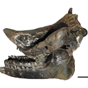
This contribution contains the 3D models described and figured in the following publication: Tissier et al. (in prep.).
Sellamynodon zimborensis UBB MPS 15795 View specimen
|
M3#297Incomplete skull with left M3. Type: "3D_surfaces"doi: 10.18563/m3.sf.297 state:published |
Download 3D surface file |
Sellamynodon zimborensis UBB MPS 15795 View specimen
|
M3#298Mandible with complete molar and premolar rows, lacking symphysis. Type: "3D_surfaces"doi: 10.18563/m3.sf.298 state:published |
Download 3D surface file |
Amynodontopsis aff. bodei UBB MPS V545 View specimen
|
M3#299Maxillary fragment with M1-3. Type: "3D_surfaces"doi: 10.18563/m3.sf.299 state:published |
Download 3D surface file |
Amynodontopsis aff. bodei UBB MPS V546 View specimen
|
M3#300Unworn m1/2 on mandible fragment. Type: "3D_surfaces"doi: 10.18563/m3.sf.300 state:published |
Download 3D surface file |
The present 3D Dataset contains the 3D models of external and internal aspects of human upper permanent second molars from the Neolithic necropolis analyzed in the following publication: Le Luyer M., Coquerelle M., Rottier S., Bayle P. (2016): Internal tooth structure and burial practices: insights into the Neolithic necropolis of Gurgy (France, 5100-4000 cal. BC). Plos One 11(7): e0159688. doi: 10.1371/journal.pone.0159688.
Homo sapiens GLN04-201-ULM2 View specimen
|
M3#74Outer enamel surface (OES) and enamel-dentine junction (EDJ) of Neolithic upper permanent left second molar Type: "3D_surfaces"doi: 10.18563/m3.sf.74 state:published |
Download 3D surface file |
Homo sapiens GLN04-206-ULM2 View specimen
|
M3#75Outer enamel surface (OES) and enamel-dentine junction (EDJ) of Neolithic upper permanent left second molar Type: "3D_surfaces"doi: 10.18563/m3.sf.75 state:published |
Download 3D surface file |
Homo sapiens GLN05-213-URM2 View specimen
|
M3#76Outer enamel surface (OES) and enamel-dentine junction (EDJ) of Neolithic upper permanent right second molar Type: "3D_surfaces"doi: 10.18563/m3.sf.76 state:published |
Download 3D surface file |
Homo sapiens GLN05-215A-URM2 View specimen
|
M3#77Outer enamel surface (OES) and enamel-dentine junction (EDJ) of Neolithic upper permanent right second molar Type: "3D_surfaces"doi: 10.18563/m3.sf.77 state:published |
Download 3D surface file |
Homo sapiens GLN06-215B-URM2 View specimen
|
M3#78Outer enamel surface (OES) and enamel-dentine junction (EDJ) of Neolithic upper permanent right second molar Type: "3D_surfaces"doi: 10.18563/m3.sf.78 state:published |
Download 3D surface file |
Homo sapiens GLN06-223-URM2 View specimen
|
M3#79Outer enamel surface (OES) and enamel-dentine junction (EDJ) of Neolithic upper permanent right second molar Type: "3D_surfaces"doi: 10.18563/m3.sf.79 state:published |
Download 3D surface file |
Homo sapiens GLN04-229-URM2 View specimen
|
M3#80Outer enamel surface (OES) and enamel-dentine junction (EDJ) of Neolithic upper permanent right second molar Type: "3D_surfaces"doi: 10.18563/m3.sf.80 state:published |
Download 3D surface file |
Homo sapiens GLN05-243B-ULM2 View specimen
|
M3#81Outer enamel surface (OES) and enamel-dentine junction (EDJ) with reconstructed dentine horn tip of Neolithic upper permanent left second molar Type: "3D_surfaces"doi: 10.18563/m3.sf.81 state:published |
Download 3D surface file |
Homo sapiens GLN04-248-ULM2 View specimen
|
M3#82Outer enamel surface (OES) and enamel-dentine junction (EDJ) with reconstructed dentine horn tip of Neolithic upper permanent left second molar Type: "3D_surfaces"doi: 10.18563/m3.sf.82 state:published |
Download 3D surface file |
Homo sapiens GLN04-252-ULM2 View specimen
|
M3#83Outer enamel surface (OES) and enamel-dentine junction (EDJ) of Neolithic upper permanent left second molar Type: "3D_surfaces"doi: 10.18563/m3.sf.83 state:published |
Download 3D surface file |
Homo sapiens GLN04-253-ULM2 View specimen
|
M3#84Outer enamel surface (OES) and enamel-dentine junction (EDJ) of Neolithic upper permanent left second molar Type: "3D_surfaces"doi: 10.18563/m3.sf.84 state:published |
Download 3D surface file |
Homo sapiens GLN05-257-URM2 View specimen
|
M3#85Outer enamel surface (OES) and enamel-dentine junction (EDJ) with reconstructed dentine horn tip of Neolithic upper permanent right second molar Type: "3D_surfaces"doi: 10.18563/m3.sf.85 state:published |
Download 3D surface file |
Homo sapiens GLN04-264-ULM2 View specimen
|
M3#86Outer enamel surface (OES) and enamel-dentine junction (EDJ) of Neolithic upper permanent left second molar Type: "3D_surfaces"doi: 10.18563/m3.sf.86 state:published |
Download 3D surface file |
Homo sapiens GLN04-277-URM2 View specimen
|
M3#87Outer enamel surface (OES) and enamel-dentine junction (EDJ) of Neolithic upper permanent right second molar Type: "3D_surfaces"doi: 10.18563/m3.sf.87 state:published |
Download 3D surface file |
Homo sapiens GLN04-289B-URM2 View specimen
|
M3#88Outer enamel surface (OES) and enamel-dentine junction (EDJ) of Neolithic upper permanent right second molar Type: "3D_surfaces"doi: 10.18563/m3.sf.88 state:published |
Download 3D surface file |
Homo sapiens GLN06-291-URM2 View specimen
|
M3#89Outer enamel surface (OES) and enamel-dentine junction (EDJ) with reconstructed dentine horn tip of Neolithic upper permanent right second molar Type: "3D_surfaces"doi: 10.18563/m3.sf.89 state:published |
Download 3D surface file |
Homo sapiens GLN05-292-URM2 View specimen
|
M3#90Outer enamel surface (OES) and enamel-dentine junction (EDJ) of Neolithic upper permanent right second molar Type: "3D_surfaces"doi: 10.18563/m3.sf.90 state:published |
Download 3D surface file |
Homo sapiens GLN05-294-ULM2 View specimen
|
M3#91Outer enamel surface (OES) and enamel-dentine junction (EDJ) with reconstructed dentine horn tip of Neolithic upper permanent left second molar Type: "3D_surfaces"doi: 10.18563/m3.sf.91 state:published |
Download 3D surface file |
Homo sapiens GLN05-308-URM2 View specimen
|
M3#93Outer enamel surface (OES) and enamel-dentine junction (EDJ) of Neolithic upper permanent right second molar Type: "3D_surfaces"doi: 10.18563/m3.sf.93 state:published |
Download 3D surface file |
Homo sapiens GLN05-301-ULM2 View specimen
|
M3#92Outer enamel surface (OES) and enamel-dentine junction (EDJ) of Neolithic upper permanent left second molar Type: "3D_surfaces"doi: 10.18563/m3.sf.92 state:published |
Download 3D surface file |
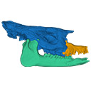
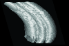
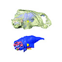
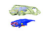
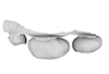
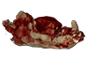
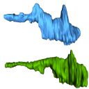
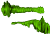
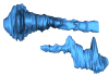
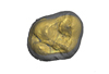
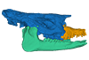
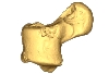
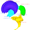
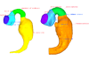
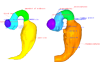
The present 3D Dataset contains two 3D models described in Tissier et al. (https://doi.org/10.1098/rsos.200633): the only known complete mandible of the early-branching rhinocerotoid Epiaceratherium magnum Uhlig, 1999, and a hypothetical reconstruction of the complete archetypic skull of Epiaceratherium Heissig, 1969, created by merging three cranial parts from three distinct Epiaceratherium species.
Epiaceratherium magnum NMB.O.B.928 View specimen

|
M3#5343D surface model of the mandible NMB.O.B.928 of Epiaceratherium magnum, with texture file. Type: "3D_surfaces"doi: 10.18563/m3.sf.534 state:published |
Download 3D surface file |
Epiaceratherium magnum NMB.O.B.928 + MJSN POI007–245 + NMB.I.O.43 View specimen
|
M3#535Archetypal reconstruction of the skull of Epiaceratherium, generated by 3D virtual association of the cranium of E. delemontense (MJSN POI007–245, in blue), mandible of E. magnum (NMB.O.B.928, green) and snout of E. bolcense (NMB.I.O.43, in orange). Type: "3D_surfaces"doi: 10.18563/m3.sf.535 state:published |
Download 3D surface file |
This contribution contains the 3D model of the fossil talus of a small-bodied anthropoid primate (Platyrrhini, Cebidae, Cebinae) discovered from lower Miocene deposits of Peruvian Amazonia (MD-61 locality, Upper Madre de Dios Basin). This fossil was described and figured in the following publication: Marivaux et al. (2012), A platyrrhine talus from the early Miocene of Peru (Amazonian Madre de Dios Sub-Andean Zone). Journal of Human Evolution. http://dx.doi.org/10.1016/j.jhevol.2012.07.005
Cebinae indet. sp. MUSM-2024 View specimen
|
M3#380Right talus 3D surface of a Miocene Cebinae indet. primate Type: "3D_surfaces"doi: 10.18563/m3.sf.380 state:published |
Download 3D surface file |
This contribution contains the 3D models described and figured in the following publication: Shiraishi N et al. Morphology and morphometry of the human embryonic brain: A three-dimensional analysis NeuroImage 115, 2015, 96-103, DOI: 10.1016/j.neuroimage.2015.04.044.
Homo sapiens KC-CS13BRN50455 View specimen

|
M3#24Computationally reconstructed cerebral parenchyma and ventricle of the human embryo at Carnegie Stage 13. Type: "3D_surfaces"doi: 10.18563/m3.sf24 state:published |
Download 3D surface file |
Homo sapiens KC-CS14BRN18834 View specimen

|
M3#25Computationally reconstructed cerebral parenchyma and ventricle of the human embryo at Carnegie Stage 14. Type: "3D_surfaces"doi: 10.18563/m3.sf25 state:published |
Download 3D surface file |
Homo sapiens KC-CS15BRN19975 View specimen
|
M3#26Computationally reconstructed cerebral parenchyma and ventricle of the human embryo at Carnegie Stage 15. Type: "3D_surfaces"doi: 10.18563/m3.sf26 state:published |
Download 3D surface file |
Homo sapiens KC-CS16BRN7870 View specimen
|
M3#27Computationally reconstructed cerebral parenchyma and ventricle of the human embryo at Carnegie Stage 16. Type: "3D_surfaces"doi: 10.18563/m3.sf27 state:published |
Download 3D surface file |
Homo sapiens KC-CS17BRN26702 View specimen

|
M3#28Computationally reconstructed cerebral parenchyma and ventricle of the human embryo at Carnegie Stage 17. Type: "3D_surfaces"doi: 10.18563/m3.sf28 state:published |
Download 3D surface file |
Homo sapiens KC-CS18BRN25914 View specimen

|
M3#29Computationally reconstructed cerebral parenchyma and ventricle of the human embryo at Carnegie Stage 18. Type: "3D_surfaces"doi: 10.18563/m3.sf29 state:published |
Download 3D surface file |
Homo sapiens KC-CS19BRN16508 View specimen

|
M3#30Computationally reconstructed cerebral parenchyma and ventricle of the human embryo at Carnegie Stage 19. Type: "3D_surfaces"doi: 10.18563/m3.sf30 state:published |
Download 3D surface file |
Homo sapiens KC-CS20BRN26581 View specimen

|
M3#31Computationally reconstructed cerebral parenchyma and ventricle of the human embryo at Carnegie Stage 20. Type: "3D_surfaces"doi: 10.18563/m3.sf31 state:published |
Download 3D surface file |
Homo sapiens KC-CS21BRN33434 View specimen

|
M3#32Computationally reconstructed cerebral parenchyma and ventricle of the human embryo at Carnegie Stage 21. Type: "3D_surfaces"doi: 10.18563/m3.sf32 state:published |
Download 3D surface file |
Homo sapiens KC-CS22BRN27960 View specimen

|
M3#33Computationally reconstructed cerebral parenchyma and ventricle of the human embryo at Carnegie Stage 22. Type: "3D_surfaces"doi: 10.18563/m3.sf33 state:published |
Download 3D surface file |
Homo sapiens KC-CS23BRN28189 View specimen

|
M3#34Computationally reconstructed cerebral parenchyma and ventricle of the human embryo at Carnegie Stage 23. Type: "3D_surfaces"doi: 10.18563/m3.sf34 state:published |
Download 3D surface file |


