Explodable 3D Dog Skull for Veterinary Education
3D models of Ocnotherium skull
3D models of Kalakocetus, the earliest Cetacea
3D GM dataset of bird skeletal variation
Skeletal embryonic development in the catshark
Bony connexions of the petrosal bone of extant hippos
bony labyrinth (14) , inner ear (11) , geometric morphometrics (10) , CT-scan (10) , Eocene (10) , Micro-CT (9) , Miocene (8)
Lionel Hautier (24) , Maëva Judith Orliac (23) , Laurent Marivaux (18) , Renaud Lebrun (14) , Rodolphe Tabuce (14) , Pierre-Olivier Antoine (13) , Bastien Mennecart (13)
|
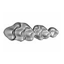
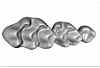
3D models related to the publication: Wild versus lab house mice: Effects of age, diet, and genetics on molar geometry and topography.Sabrina Renaud
Published online: 06/08/2021 |

|
M3#736BW_03 Type: "3D_surfaces"doi: 10.18563/m3.sf.736 state:published |
Download 3D surface file |
Mus musculus BW_04 View specimen

|
M3#752BW_04 Type: "3D_surfaces"doi: 10.18563/m3.sf.752 state:published |
Download 3D surface file |
Mus musculus BW_06 View specimen

|
M3#753BW_06 Type: "3D_surfaces"doi: 10.18563/m3.sf.753 state:published |
Download 3D surface file |
Mus musculus BW_07 View specimen

|
M3#754BW_07 Type: "3D_surfaces"doi: 10.18563/m3.sf.754 state:published |
Download 3D surface file |
Mus musculus BW_08 View specimen

|
M3#755BW_08 Type: "3D_surfaces"doi: 10.18563/m3.sf.755 state:published |
Download 3D surface file |
Mus musculus BW_11 View specimen

|
M3#756BW_11 Type: "3D_surfaces"doi: 10.18563/m3.sf.756 state:published |
Download 3D surface file |
Mus musculus BW_12 View specimen

|
M3#757BW_12 Type: "3D_surfaces"doi: 10.18563/m3.sf.757 state:published |
Download 3D surface file |
Mus musculus Blab_035 View specimen

|
M3#758Blab_035 Type: "3D_surfaces"doi: 10.18563/m3.sf.758 state:published |
Download 3D surface file |
Mus musculus Blab_046 View specimen

|
M3#759Blab_046 Type: "3D_surfaces"doi: 10.18563/m3.sf.759 state:published |
Download 3D surface file |
Mus musculus Blab_054 View specimen

|
M3#760Blab_054 Type: "3D_surfaces"doi: 10.18563/m3.sf.760 state:published |
Download 3D surface file |
Mus musculus Blab_056 View specimen

|
M3#761Blab_056 Type: "3D_surfaces"doi: 10.18563/m3.sf.761 state:published |
Download 3D surface file |
Mus musculus Blab_082 View specimen

|
M3#762Blab_082 Type: "3D_surfaces"doi: 10.18563/m3.sf.762 state:published |
Download 3D surface file |
Mus musculus Blab_086 View specimen

|
M3#763Blab_086 Type: "3D_surfaces"doi: 10.18563/m3.sf.763 state:published |
Download 3D surface file |
Mus musculus Blab_092 View specimen

|
M3#764Blab_092 Type: "3D_surfaces"doi: 10.18563/m3.sf.764 state:published |
Download 3D surface file |
Mus musculus Blab_319 View specimen

|
M3#751Blab_319 Type: "3D_surfaces"doi: 10.18563/m3.sf.751 state:published |
Download 3D surface file |
Mus musculus Blab_325 View specimen

|
M3#750Blab_325 Type: "3D_surfaces"doi: 10.18563/m3.sf.750 state:published |
Download 3D surface file |
Mus musculus Blab_329 View specimen

|
M3#737Blab_329 Type: "3D_surfaces"doi: 10.18563/m3.sf.737 state:published |
Download 3D surface file |
Mus musculus Blab_330 View specimen

|
M3#738Blab_330 Type: "3D_surfaces"doi: 10.18563/m3.sf.738 state:published |
Download 3D surface file |
Mus musculus Blab_F2a View specimen

|
M3#739Blab_F2a Type: "3D_surfaces"doi: 10.18563/m3.sf.739 state:published |
Download 3D surface file |
Mus musculus Blab_F2b View specimen

|
M3#740Blab_F2b Type: "3D_surfaces"doi: 10.18563/m3.sf.740 state:published |
Download 3D surface file |
Mus musculus Blab_BB3w View specimen

|
M3#741Blab_BB3w Type: "3D_surfaces"doi: 10.18563/m3.sf.741 state:published |
Download 3D surface file |
Mus musculus hyb_BS01 View specimen

|
M3#742hyb_BS01 Type: "3D_surfaces"doi: 10.18563/m3.sf.742 state:published |
Download 3D surface file |
Mus musculus hyb_BS02 View specimen
|
M3#743hyb_BS02 Type: "3D_surfaces"doi: 10.18563/m3.sf.743 state:published |
Download 3D surface file |
Mus musculus hyb_SB01 View specimen

|
M3#744hyb_SB01 Type: "3D_surfaces"doi: 10.18563/m3.sf.744 state:published |
Download 3D surface file |
Mus musculus hyb_SB02 View specimen

|
M3#745hyb_SB02 Type: "3D_surfaces"doi: 10.18563/m3.sf.745 state:published |
Download 3D surface file |
Mus musculus SW_001 View specimen

|
M3#746SW_001 Type: "3D_surfaces"doi: 10.18563/m3.sf.746 state:published |
Download 3D surface file |
Mus musculus SW_002 View specimen

|
M3#747SW_002 Type: "3D_surfaces"doi: 10.18563/m3.sf.747 state:published |
Download 3D surface file |
Mus musculus SW_005 View specimen

|
M3#748SW_005 Type: "3D_surfaces"doi: 10.18563/m3.sf.748 state:published |
Download 3D surface file |
Mus musculus SW_0ter View specimen

|
M3#749SW_0ter Type: "3D_surfaces"doi: 10.18563/m3.sf.749 state:published |
Download 3D surface file |
Mus musculus SW_343 View specimen

|
M3#765SW_343 Type: "3D_surfaces"doi: 10.18563/m3.sf.765 state:published |
Download 3D surface file |
This contribution contains the 3D model of the holotype of Simplomys hugi, the new dormouse species from the locality of Glovelier described and figured in the following publication: New data on the Miocene dormouse Simplomys García-Paredes, 2009 from the peri-alpin basins of Switzerland and Germany: palaeodiversity of a rare genus in Central Europe. https://doi.org/10.1007/s12549-018-0339-y
Simplomys hugi MJSN-GLM017-0001 View specimen
|
M3#385the left maxilla with four teeth ( DP4, P4, M1 and M2) Type: "3D_surfaces"doi: 10.18563/m3.sf.385 state:published |
Download 3D surface file |
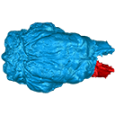
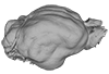
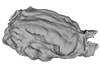
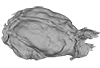
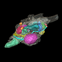
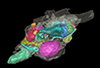
The present 3D Dataset contains the 3D models analyzed in: Amson et al., Under review. Evolutionary Adaptation to Aquatic Lifestyle in Extinct Sloths Can Lead to Systemic Alteration of Bone Structure doi:10.1098/rspb.2018.0270.
Bradypus tridactylus MNHN ZM-MO-1999-1065 View specimen

|
M3#337Brain endocast Type: "3D_surfaces"doi: 10.18563/m3.sf.337 state:published |
Download 3D surface file |
Choloepus didactylus MNHN-ZM-MO-1996-594 View specimen
|
M3#338Brain endocast Type: "3D_surfaces"doi: 10.18563/m3.sf.338 state:published |
Download 3D surface file |
Thalassocnus natans MNHN-F-SAS-734 View specimen

|
M3#339Brain endocast Type: "3D_surfaces"doi: 10.18563/m3.sf.339 state:published |
Download 3D surface file |
Thalassocnus littoralis MNHN-F-SAS-1610 View specimen
|
M3#340Brain endocast Type: "3D_surfaces"doi: 10.18563/m3.sf.340 state:published |
Download 3D surface file |
Thalassocnus littoralis MNHN-F-SAS-1615 View specimen
|
M3#341Brain endocast Type: "3D_surfaces"doi: 10.18563/m3.sf.341 state:published |
Download 3D surface file |
Thalassocnus carolomartini SMNK-3814 View specimen
|
M3#342Brain endocast lacking right olfactory bulb Type: "3D_surfaces"doi: 10.18563/m3.sf.342 state:published |
Download 3D surface file |
The present Dataset contains the 3D model of the male genital organs of greater horseshoe bat, Rhinolophus ferrumequinum. This is the first detailed 3D structure of the soft-tissue genital organs of bats. The 3D model was generated using microCT and techniques of virtual reconstruction.
Rhinolophus ferrumequinum JP18-006 View specimen
|
M3#521The genital organs of male greater horseshoe bat. Type: "3D_surfaces"doi: 10.18563/m3.sf.521 state:published |
Download 3D surface file |
This contribution contains the 3D models described and figured in the following publication: Orliac M.J., Karadenizli L., Antoine P.-O., Sen S. 2015. Small suids (Mammalia, Artiodactyla) from the late Early Miocene of Turkey and a short overview of Early Miocene small suoids in the Old World. Paleontologia electronica 18(2): 18.2.30A: 1-48. https://doi.org/10.26879/547
?Nguruwe galaticum SMT-1 View specimen
|
M3#16fragment of palate with left broken M1-M3 Type: "3D_surfaces"doi: 10.18563/m3.sf16 state:published |
Download 3D surface file |
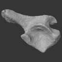
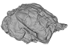
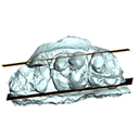
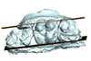
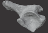
The present 3D Dataset contains the 3D models of an ilium, a vertebra, and a partial scapula of Prestosuchus sp. that were analyzed in “New Loricata remains from the Pinheiros-Chiniquá Sequence (Middle-Upper Triassic), southern Brazil”.
Prestosuchus sp. UFSM11603 View specimen
|
M3#1080Surface scan of a right ilium of Prestosuchus sp. with a 0.4 mm resolution. Type: "3D_surfaces"doi: 10.18563/m3.sf.1080 state:published |
Download 3D surface file |
Prestosuchus sp. UFSM11233 View specimen
|
M3#1081Surface scan of a partial right scapula of Prestosuchus sp. with a 0.4mm resolution. Type: "3D_surfaces"doi: 10.18563/m3.sf.1081 state:published |
Download 3D surface file |
Prestosuchus sp. UFSM11602a View specimen
|
M3#1082Surface scan of a anterior dorsal vertebra of Prestosuchus sp. with a 0.2 mm resolution. Type: "3D_surfaces"doi: 10.18563/m3.sf.1082 state:published |
Download 3D surface file |


